. 2006 Mar;72(3):1900-9.
doi: 10.1128/AEM.72.3.1900-1909.2006.

Chromosomal dynamism in progeny of outbreak-related sorbitol-fermenting enterohemorrhagic Escherichia coli O157:NM

Affiliations

Chromosomal dynamism in progeny of outbreak-related sorbitol-fermenting enterohemorrhagic Escherichia coli O157:NM

Martina Bielaszewska et al. Appl Environ Microbiol. 2006 Mar.

Abstract

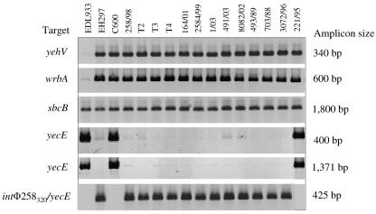
Sorbitol-fermenting (SF) enterohemorrhagic Escherichia coli (EHEC) O157:NM (nonmotile) is a unique clone that causes outbreaks of hemorrhagic colitis and hemolytic-uremic syndrome. In well-defined clusters of cases, we have observed significant variability in pulsed-field gel electrophoresis (PFGE) patterns which could indicate coinfection by different strains. An analysis of randomly selected progeny colonies of an outbreak strain after subcultivation demonstrated that they displayed either the cognate PFGE outbreak pattern or one of four additional patterns and were <89% similar. These profound alterations were associated with changes in the genomic position of one of two Shiga toxin 2-encoding genes (stx2) in the outbreak strain or with the loss of this gene. The two stx2 alleles in the outbreak strain were identical but were flanked with phage-related sequences with only 77% sequence identity. Neither of these phages produced plaques, but one lysogenized E. coli K-12 and integrated in yecE in the lysogens and the wild-type strain. The presence of two stx2 genes which correlated with increased production of Stx2 in vitro but not with the clinical outcome of infection was also found in 14 (21%) of 67 SF EHEC O157:NM isolates from sporadic cases of human disease. The variability of PFGE patterns for the progeny of a single colony must be considered when interpreting PFGE patterns in SF EHEC O157-associated outbreaks.


Figures

FIG. 1.
FIG. 1.
PFGE of XbaI-digested genomic DNAs (A) and Southern blot hybridization with an stxA2 probe (B) of SF EHEC O157:NM strains isolated during a family outbreak from a patient with HUS (strain 258/98; lanes 1), his brother with diarrhea (strain 269/98; lanes 2), and an epidemiologically associated cow (strain 550/98; lanes 3). Lanes S, molecular size standard (E. coli O157:H7 strain G5244; CDC). The sizes of XbaI fragments that hybridized with the stxA2 probe are indicated in panel B. Designations of PFGE and stxA2 hybridization patterns are given below the figure.
FIG. 2.
FIG. 2.
PFGE of XbaI-digested genomic DNAs (A) and stxA2-specific Southern blot hybridization (B) of representative progenies of strain 258/98. Lanes S, molecular size standard (E. coli O157:H7 strain G5244; CDC). In lanes 1 to 5, the following progeny colonies (genomic positions of stx2 on XbaI fragments [sizes of fragments are given in kilobases] are given in parentheses) are shown: lanes 1, progeny 3 (450 and 320); lanes 2, progeny 7 (490 and 320); lanes 3, progeny 1 (650 and 320); lanes 4, progeny 9 (390 and 320); and lanes 5, progeny 2 (320). The XbaI fragments which hybridized with the stxA2 probe are indicated in panel A, and their sizes are given in panel B. Designations of PFGE and stxA2 hybridization patterns and the numbers of progenies that displayed the respective patterns are indicated below the figure.
FIG. 3.
FIG. 3.
Cluster analysis of PFGE patterns of representative progenies of strain 258/98 which harbored stx2 on different XbaI genomic fragments or had lost one stx2 copy. (A) PFGE gel; (B) dendrogram derived from the PFGE data with BioNumerics software. The analysis of the bands generated was performed using the Dice coefficient and the unweighted-pair group method using average linkages. Lanes S, molecular size standard (S. enterica serovar Braenderup strain H9812; CDC). The progeny colonies investigated (P) and their PFGE pattern designations (A to E) are indicated. The bands which hybridized with the stxA2 probe are shown on the PFGE gel.
FIG. 4.
FIG. 4.
PFGE of XbaI-digested genomic DNAs (A) and stxA2-specific Southern blot hybridization (B) of representative SF EHEC O157:NM strains which contain one or two stx2 genes in different genomic positions and of lysogens of strain 258/98. Lanes S, molecular size standard (E. coli O157:H7 strain G5244; CDC). The following strains (stx2 positions on XbaI fragments [sizes of fragments are given in kilobases] are given in parentheses) are depicted in each panel: lanes 1, 164/01 (490 and 320); lanes 2, 25/99 (450 and 320); lanes 3, 2584/99 (490 and 210); lanes 4, 1/03 (490 and 170); lanes 5, 491/03 (370 and 320); lanes 6, 8082/02 (310 and 300); lanes 7, 493/89 (320); lanes 8, 221/95 (300); lanes 9, 703/88 (210); lanes 10, 3072/96 (150); lanes 11, lysogen T2 (260); lanes 12, lysogen T3 (260); and lanes 13, lysogen T4 (260). The sizes of the XbaI fragments that hybridized with the stxA2 probe are indicated in panel B.
FIG. 5.
FIG. 5.
Structures of stxB2-flanking regions of two stx2 genes located on the 320-kb and 450-kb XbaI fragments of strain 258/98. Analyzed sequences and their lengths are given on the left side. Arrows indicate the lengths and directions of ORFs. Significant nucleotide sequence identities between the two 258/98 stxB2-flanking regions and their identities with the most closely related sequences in GenBank are shown in light shaded boxes. The overall nucleotide sequence identity between the two stxB2-flanking regions of strain 258/98 is shown in a hatched box. Bar, 1 kb.
FIG. 6.
FIG. 6.
PCR analyses of phage integration sites in SF EHEC O157:NM. The strains tested, loci examined, and lengths of resulting amplicons are listed across the top and to the left and right of the rows of amplicons, respectively. Primers for the respective PCRs are listed in Table 2. Strains EDL933 (with stx1- and stx2-converting phages integrated in yehV and wrbA, respectively) (39, 40), EH297 (with a stx2-converting phage integrated in yecE) (9), and E. coli K-12 C600 (all of the genes investigated as putative phage integration sites are intact) (7) were used as controls.

References

    1. Ammon, A., L. R. Peterson, and H. Karch. 1999. A large outbreak of hemolytic uremic syndrome caused by an unusual sorbitol-fermenting strain of E. coli O157:H−. J. Infect. Dis. 179:1274-1277. - PubMed
    1. Barrett, T. J., H. Lior, J. H. Green, R. Khakhria, and J. G. Wells. 1994. Laboratory investigation of a multistate food-borne outbreak of Escherichia coli O157:H7 by using pulsed-field gel electrophoresis and phage typing. J. Clin. Microbiol. 32:3013-3017. - PMC - PubMed
    1. Bettelheim, K. A., M. Whipp, S. P. Djordjevic, and V. Ramachandran. 2002. First isolation outside Europe of sorbitol-fermenting verocytotoxigenic Escherichia coli (VTEC) belonging to O group O157. J. Med. Microbiol. 51:713-714. - PubMed
    1. Bielaszewska, M., H. Schmidt, A. Liesegang, R. Prager, W. Rabsch, H. Tschäpe, A. Cizek, J. Janda, K. Blahova, and H. Karch. 2000. Cattle can be a reservoir of sorbitol-fermenting Shiga toxin-producing Escherichia coli O157:H− strains and a source of human diseases. J. Clin. Microbiol. 38:3470-3473. - PMC - PubMed
    1. Bielaszewska, M., and H. Karch. 2005. Consequences of enterohaemorrhagic Escherichia coli infection for the vascular endothelium. Thromb. Haemost. 94:312-318. - PubMed

Publication types

MeSH terms

LinkOut - more resources