. 2006 Mar;72(3):2070-9.
doi: 10.1128/AEM.72.3.2070-2079.2006.

The atu and liu clusters are involved in the catabolic pathways for acyclic monoterpenes and leucine in Pseudomonas aeruginosa

Affiliations

The atu and liu clusters are involved in the catabolic pathways for acyclic monoterpenes and leucine in Pseudomonas aeruginosa

J A Aguilar et al. Appl Environ Microbiol. 2006 Mar.

Abstract

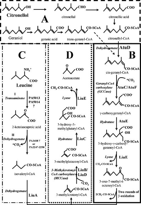
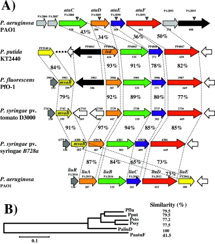
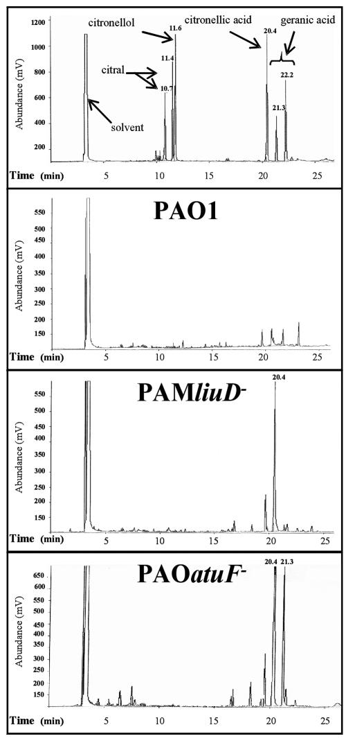
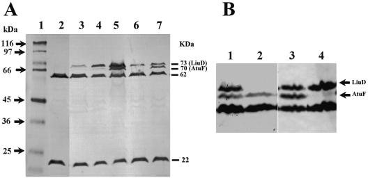
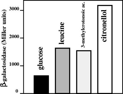
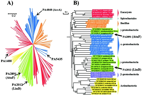
Evidence suggests that the Pseudomonas aeruginosa PAO1 gnyRDBHAL cluster, which is involved in acyclic isoprenoid degradation (A. L. Díaz-Pérez, N. A. Zavala-Hernández, C. Cervantes, and J. Campos-García, Appl. Environ. Microbiol. 70:5102-5110, 2004), corresponds to the liuRABCDE cluster (B. Hoschle, V. Gnau, and D. Jendrossek, Microbiology 151:3649-3656, 2005). A liu (leucine and isovalerate utilization) homolog cluster was found in the PAO1 genome and is related to the catabolism of acyclic monoterpenes of the citronellol family (AMTC); it was named the atu cluster (acyclic terpene utilization), consisting of the atuCDEF genes and lacking the hydroxymethyl-glutaryl-coenzyme A (CoA) lyase (HMG-CoA lyase) homolog. Mutagenesis of the atu and liu clusters showed that both are involved in AMTC and leucine catabolism by encoding the enzymes related to the geranyl-CoA and the 3-methylcrotonyl-CoA pathways, respectively. Intermediary metabolites of the acyclic monoterpene pathway, citronellic and geranic acids, were accumulated, and leucine degradation rates were affected in both atuF and liuD mutants. The alpha subunit of geranyl-CoA carboxylase and the alpha subunit of 3-methylcrotonyl-CoA carboxylase (alpha-MCCase), encoded by the atuF and liuD genes, respectively, were both induced by citronellol, whereas only the alpha-MCCase subunit was induced by leucine. Both citronellol and leucine also induced a LacZ transcriptional fusion at the liuB gene. The liuE gene encodes a probable hydroxy-acyl-CoA lyase (probably HMG-CoA lyase), an enzyme with bifunctional activity that is essential for both AMTC and leucine degradation. P. aeruginosa PAO1 products encoded by the liuABCD cluster showed a higher sequence similarity (77.2 to 79.5%) with the probable products of liu clusters from several Pseudomonas species than with the atuCDEF cluster from PAO1 (41.5%). Phylogenetic studies suggest that the atu cluster from P. aeruginosa could be the result of horizontal transfer from Alphaproteobacteria. Our results suggest that the atu and liu clusters are bifunctional operons involved in both the AMTC and leucine catabolic pathways.


Figures

FIG. 1.
FIG. 1.
Catabolic pathways and proposed functions of the atu and liu cluster products from P. aeruginosa PAO1. (A and B) Upper and lower pathways of acyclic monoterpene catabolism; (C and D) Upper and lower pathways of leucine catabolism. AtuD, citronellyl-CoA dehydrogenase; AtuC/AtuF, geranyl-CoA carboxylase; AtuE, γ-carboxygeranyl-CoA hydratase; LiuE, 3-hydroxy-γ-carboxygeranyl-CoA and 3-hydroxy-3-methylglutaryl-CoA lyase; I, leucine transaminase; II, 2-ketoisocaproic dehydrogenase; LiuA, isovaleryl-CoA dehydrogenase; LiuB/LiuD, 3-methylcrotonyl- CoA carboxylase; LiuC, 3-methylglutaconyl-CoA hydratase. (Adapted from references , , , and .)
FIG. 2.
FIG. 2.
Sequence analysis of products encoded in the atu and liu gene clusters. (A) Genetic arrangement of the atu/liu homolog clusters from Pseudomonas species. The species and strain names are shown at the left. ORF arrangement and transcription direction are shown by arrows. Locations of transposon insertions in the P. aeruginosa mutants are indicated with shaded arrowheads. Numbers above arrows show the ORF number assigned in the respective genome sequencing project. Identified genes are also shown. The amino acid number of the ORF is indicated below the arrows. Corresponding ortholog proteins are indicated by the same color. Orange, LiuA; green, LiuB; blue, LiuC; red, LiuD; yellow, lyase (LiuE); black, PA2893.The percentages of amino acid identity between upper and lower ORFs are indicated. (B) Phylogenetic tree of the Atu/Liu homolog proteins of Pseudomonas species. The amino acid sequences of LiuABCD and AtuDCEF were put together as one sequence and aligned with the homologous sequences from the strains indicated. The alignment was done using the Clustal W software, and the tree was done by the neighbor joining in the Mega2 software with a bootstrap of 1,000 replicates. The numbers indicate the percent similarities between the amino acids sequences aligned. Pflu, P. fluorescens PfO-1; Pput, P. putida KT2440; Psto, P. syringae pv. tomato D3000; Pssy, P. syringae pv. syringae B728a; PaatuF, P. aeruginosa PAO1 atu cluster; PaliuD, P. aeruginosa PAO1 liu cluster. The evolutionary distance scale bar is shown at the bottom.
FIG. 3.
FIG. 3.
Metabolite accumulation in cultures of P. aeruginosa grown in citronellol. Cultures were grown in M9 medium with succinic acid as the carbon source for 18 h, and citronellol was then added. The metabolites were extracted from the supernatants and analyzed at 48 h after the addition of citronellol by gas chromatography as described in Materials and Methods. Commercial citral (10.7 and 11.4 min), citronellol (11.6 min), citronellic acid (20.4 min), and geranic acid mix isomers (21.3 and 22.2 min) were used as standard compounds. Peaks with retention times of 20.4 and 21.3 min correspond to citronellic acid and geranic acid, respectively.
FIG. 4.
FIG. 4.
Western blot analysis of biotinylated proteins from P. aeruginosa PAO1 derivatives. (A) Lanes: 1, molecular masses of protein standards; 2 to 7, cell extracts from the PAO1 strain grown on glucose (lane 2), isoleucine (lane 3), leucine (lane 4), citronellol (lane 5), glucose plus leucine (lane 6), and glucose plus citronellol (lane 7). Proteins of 73, 70, 62, and 22 kDa identified with avidin-HRP conjugate, corresponding to biotinylated subunits from alpha subunits of geranyl-CoA carboxylase (AtuF), 3-methylcrotonyl-CoA carboxylase (LiuD), a putative acyl-CoA carboxylase, and acetyl-CoA carboxylase, respectively, are indicated. (B) Biotinylated proteins from cultures grown in glucose plus citronellol. Lanes: 1 and 3, PAO1 wild-type strain; 2, PAM liuD mutant; 4, PAO atuF mutant. The positions of the LiuD and AtuF proteins are indicated.
FIG. 5.
FIG. 5.
Induction of the liuB::lacZ transcriptional fusion. Cultures of the PAO1 strain with plasmid pANP2 (P2liuB::lacZ) were grown in M9 medium with glucose. β-Galactosidase activity was measured after the addition of the inducer compounds glucose (0.2%), leucine (0.005%), 3-methylcrotonoic acid (0.005%), and citronellol (0.005%). Data given correspond to Miller units after 3 h of induction with the compound indicated and are the averages of two independent experiments done in duplicate; the standard deviation was less than 5% of the given value.
FIG. 6.
FIG. 6.
Phylogenetic trees of AtuF/LiuD protein orthologs. Trees were obtained by the UPGMA, NJ, and ME methods in the Mega2 package as described in Materials and Methods, and the NJ tree is shown. (A) The tree shows the five phylogenetic groups of the biotinylated subunits of acyl-CoA carboxylases aligned; the probable functions of the ortholog proteins are indicated in the phylogenetic groups, in agreement with some characterized carboxylases: acetyl-CoA carboxylases (blue), propionyl-CoA carboxylases (orange), 3-methylcrotonyl-CoA carboxylases (green), pyruvate carboxylases (red), and urea carboxylases (yellow). Acyl-CoA carboxylases from P. aeruginosa PAO1 are shown by arrows. (B) Phylogenetic tree of MCCase root constructed with the 41 proteins showing the higher phylogenetic relationship. The AtuF and LiuD proteins of P. aeruginosa are shown by arrows. Bootstrap values of higher than 50% obtained by the UPGMA, NJ, and ME methods are shown. Blue, Alphaproteobacteria; purple, Betaproteobacteria; green, Gammaproteobacteria; yellow, Actinobacteria; orange, Bacillus; gray, Spirochaeta; red, eukaryote.

References

    1. Anderson, M. D., P. Che, J. Song, B. J. Nikolau, and E. S. Wurtele. 1998. 3-Methylcrotonyl-coenzyme A carboxylase is a component of the mitochondrial leucine catabolic pathway in plants. Plant Physiol. 118:1127-1138. - PMC - PubMed
    1. Campos-García, J., and G. Soberón-Chávez. 2000. Degradation of the methyl substituted alkene, citronellol, by Pseudomonas aeruginosa, wild type and mutant strains. Biotechnol. Lett. 22:235-237.
    1. Cantwell, S. G., E. P. Lau, D. S. Watt, and R. R. Fall. 1978. Biodegradation of acyclic isoprenoids by Pseudomonas species. J. Bacteriol. 153:324-333. - PMC - PubMed
    1. Conrad, R. S., L. K. Massey, and J. R. Sokatch. 1974. d- and l-isoleucine metabolism and regulation of their pathways in Pseudomonas putida. J. Bacteriol. 11:103-111. - PMC - PubMed
    1. Díaz-Pérez, A. L., N. A. Zavala-Hernández, C. Cervantes, and J. Campos-García. 2004. The atuRDBHAL cluster is involved in acyclic isoprenoid degradation in Pseudomonas aeruginosa. Appl. Environ. Microbiol. 70:5102-5110. - PMC - PubMed

Publication types