. 2006 Feb;2(2):e14.
doi: 10.1371/journal.ppat.0020014. Epub 2006 Feb 24.

Downregulation of the Drosophila immune response by peptidoglycan-recognition proteins SC1 and SC2

Affiliations

Downregulation of the Drosophila immune response by peptidoglycan-recognition proteins SC1 and SC2

Vincent Bischoff et al. PLoS Pathog. 2006 Feb.

Abstract

Peptidoglycan-recognition proteins (PGRPs) are evolutionarily conserved molecules that are structurally related to bacterial amidases. Several Drosophila PGRPs have lost this enzymatic activity and serve as microbe sensors through peptidoglycan recognition. Other PGRP family members, such as Drosophila PGRP-SC1 or mammalian PGRP-L, have conserved the amidase function and are able to cleave peptidoglycan in vitro. However, the contribution of these amidase PGRPs to host defense in vivo has remained elusive so far. Using an RNA-interference approach, we addressed the function of two PGRPs with amidase activity in the Drosophila immune response. We observed that PGRP-SC1/2-depleted flies present a specific over-activation of the IMD (immune deficiency) signaling pathway after bacterial challenge. Our data suggest that these proteins act in the larval gut to prevent activation of this pathway following bacterial ingestion. We further show that a strict control of IMD-pathway activation is essential to prevent bacteria-induced developmental defects and larval death.


Conflict of interest statement

Competing interests. The authors have declared that no competing interests exist.

Figures

Figure 1
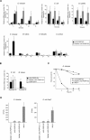
Figure 1. Specific Reduction of PGRP-SC1/2 mRNA Using RNA Interference In Vivo
Each histogram corresponds to the mean value of four independent experiments (± standard deviation). (A) mRNA-level quantification for different PGRPs (PGRP/RpL32) shows that PGRP-SC1 and PGRP-SC2 mRNA levels are severely reduced in DaGal4;UAS iPGRP-SC flies as compared to UAS iPGRP-SC control flies. PGRP-SA and PGRP-SD mRNA levels are not affected. One hundred percent corresponds to the wild-type value for each transcript. Asterisks indicate that the difference between UAS iPGRP-SC and DaGal4;UAS iPGRP-SC values is statistically significant (p < 0.05). (B) Primer specificity in RT-PCR experiments shown by quantification of PGRP-SC1b and PGRP-SC2 transcripts. PGRP-SC1b over-expression in HspGal4;UAS PGRP-SC1b flies 1 h after a 30-min heat-shock (37 °C) treatment (HS) is well detected with PGRP-SC1 primers but not with those for PGRP-SC2. We can infer that the PGRP-SC2 primers used in this study are able to discriminate between PGRP-SC2 and PGRP-SC1b transcripts.
Figure 2
Figure 2. IMD-Pathway Activation Is Downregulated by PGRP-SC1/2
(A) Kinetics of diptericin mrna induction (Dipt/RpL32) after infection by various bacteria. Each histogram corresponds to the mean value of five independent experiments (± standard deviation). Asterisks indicate that the difference between DaGal4;UAS iPGRP-SC and control UAS iPGRP-SC values is statistically significant (p < 0.05). One hundred percent corresponds to the level of activation at 6 h in control flies. In the lower panel, diptericin induction after gram-positive bacterial infections were compared to that obtained after infection by E. cloacae (100%). RpL32 is used as an internal control. ci, clean injury; ni, noninfected. (B) Quantification of diptericin mRNA levels in UAS iPGRP-SA, DaGal4;UAS iPGRP-SA and PGRP-SAseml flies shows that reduction of PGRP-SA mRNA levels does not influence IMD-pathway induction 6 h after infection by E. coli. Quantification of drosomycin mRNA levels 24 h after M. luteus infection indicates that PGRP-SA is efficiently knocked down by dsRNA interference. PGRP-SAseml is a complete loss-of-function mutant for PGRP-SA. Each histogram corresponds to the mean value of five independent experiments (± standard deviation). Asterisk indicates that the difference between DaGal4;UAS iPGRP-SA and control UAS iPGRP-SA values is statistically significant (p < 0.05). (C) DaGal4;UAS iPGRP-SC flies are as susceptible to infection by E. cloacae as control flies. (D) E. cloacae and E. coli AmpR growth in various genetic backgrounds 24 h after infection. Flies with reduced levels of PGRP-SC1/2, unlike IKKγkey1 mutants, are able to clear bacteria from their hemolymph. Each histogram corresponds to the mean value of four independent experiments (± standard deviation).
Figure 3
Figure 3. Toll-Pathway Activation Is Wild-Type in DaGal4;UAS iPGRP-SC Flies
Kinetics of drosomycin mrna induction (Drs/RpL32) after infection by gram-positive (upper panel) and gram-negative (lower panel) bacteria. Each histogram corresponds to the mean value of six independent experiments (± standard deviation). Asterisk indicates that the difference between DaGal4;UAS iPGRP-SC and control UAS iPGRP-SC values is statistically significant (p < 0.05). One hundred percent corresponds to the level of activation 24 h after infection in control flies. In the lower panel, drosomycin induction after gram-negative bacterial infections is compared to that of S. aureus infection which is set to 100%. RpL32 is used as an internal control.
Figure 4
Figure 4. Reduction of PGRP-SC1/2 Levels in the Larval Gut Increases IMD-Pathway Activation after Natural Infection
(A) PGRP-SC1 and PGRP-SC2 mRNAs (PGRP-SC/RpL32) are mainly expressed in the larval gut and are severely reduced in DaGal4;UAS iPGRP-SC and CadGal4;UAS iPGRP-SC larvae. Each histogram corresponds to the mean value of four independent experiments (± standard deviation). (B) Diptericin mrna induction levels (Dipt/RpL32), measured 6 h and 24 h after natural infection. Each histogram corresponds to the mean value of variable numbers (shown in parentheses) of independent experiments (± standard deviation). Asterisks indicate that the difference between DaGal4;UAS iPGRP-SC or CadGal4;UAS iPGRP-SC and control UAS iPGRP-SC values is statistically significant (p < 0.05). (C) Drosomycin mrna induction levels (Drs/RpL32), measured 24 h after natural infection. Each histogram corresponds to the mean value of four independent experiments (± standard deviation). (D) Three hours after natural infection with E. coli GFP, bacteria were found to be highly concentrated in the anterior half of the larval gut. In larvae with reduced gut PGRP-SC levels (CadGal4;UAS iPGRP-SC), feeding on E.coli is sufficient to trigger IMD-pathway activation in the fat body after 24 h (visualized here by the use of a diptericin-LacZ transgene). (E) Percentage of larvae showing β-galactosidase activity in the fat body 24 h after natural infection. For each genotype, ten larvae were dissected and stained. Each histogram corresponds to the mean value of five independent experiments (± standard deviation). Asterisks indicate that the difference between diptLacZ;UAS iPGRP-SC and diptLacZ;DaGal4;UAS iPGRP-SC or diptLacZ;CadGal4;UAS iPGRP-SC values and between diptLacZ,UAS iPGRP-SC and diptLacZ;CadGal4;UAS iPGRP-SC values is statistically significant (p < 0.05).
Figure 5
Figure 5. Reduction of PGRP-SC1/2 Levels Sensitizes Larvae to Bacterial Infection
(A) The percentage of dead larvae is measured 24 h after natural infection. Numbers in parentheses correspond to the total number of infected larvae. Each histogram corresponds to the mean value of six independent experiments (± standard deviation) for E. coli and five for E. carotovora carotovora. Asterisks indicate that the difference between UAS iPGRP-SC and DaGal4;UAS iPGRP-SC values is statistically significant (p < 0.05). (B) The percentage of adults showing wing notching is measured 7 d after natural infection. Numbers in parentheses correspond to the total number of infected larvae. (C–F) Natural infection with E. carotovora carotorova and E. coli triggers increased cell death and developmental defects in DaGal4;UAS iPGRP-SC flies. Wing imaginal discs dissected from DaGal4;UAS iPGRP-SC larvae (F) show higher levels of cell death after natural infection than discs from infected UAS iPGRP-SC control larvae (E). Consistently, some DaGal4;UAS iPGRP-SC adults derived from infected larvae exhibit wing notching (indicated by arrowheads) (D), which was never observed in infected controls (C).

References

    1. Bulet P, Charlet M, Hetru C. Antimicrobial peptides in insect immunity. In: Ezekowitz RAB, Hoffmann JA, editors. Infectious disease: Innate immunity. Totowa (New Jersey): Human Press; 2002. pp. 89–107.
    1. Zasloff M. Antimicrobial peptides of multicellular organisms. Nature. 2002;415:389–395. - PubMed
    1. De Gregorio E, Spellman PT, Rubin GM, Lemaitre B. Genome-wide analysis of the Drosophila immune response by using oligonucleotide microarrays. Proc Natl Acad Sci U S A. 2001;98:12590–12595. - PMC - PubMed
    1. Irving P, Troxler L, Heuer TS, Belvin M, Kopczynski C, et al. A genome-wide analysis of immune responses in Drosophila . Proc Natl Acad Sci U S A. 2001;98:15119–15124. - PMC - PubMed
    1. Hoffmann JA. The immune response of Drosophila . Nature. 2003;426:33–38. - PubMed

Publication types

Substances