Review
. 2006 Feb;23(4):921-34.
doi: 10.1111/j.1460-9568.2006.04611.x.

Comparative aspects of cerebral cortical development

Affiliations
Review

Comparative aspects of cerebral cortical development

Zoltán Molnár et al. Eur J Neurosci. 2006 Feb.

Abstract

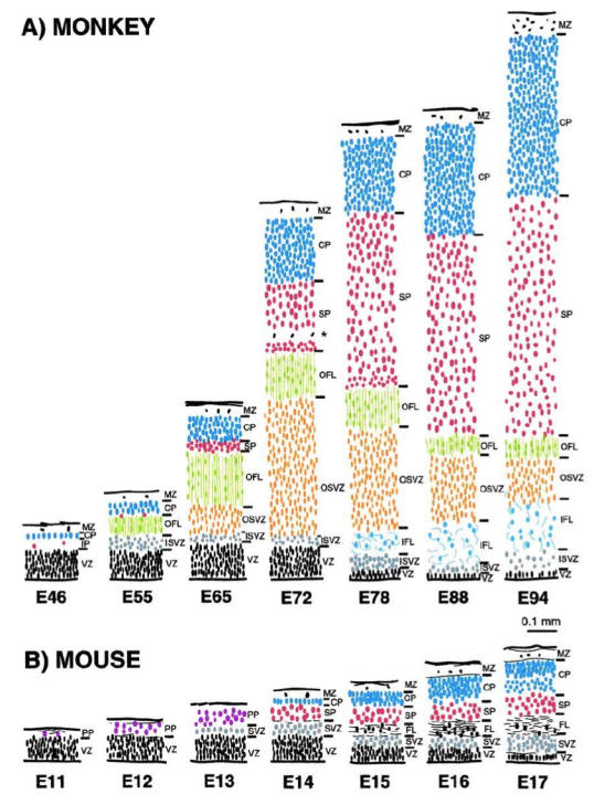
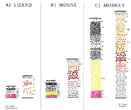
This review aims to provide examples of how both comparative and genetic analyses contribute to our understanding of the rules for cortical development and evolution. Genetic studies have helped us to realize the evolutionary rules of telencephalic organization in vertebrates. The control of the establishment of conserved telencephalic subdivisions and the formation of boundaries between these subdivisions has been examined and the very specific alterations at the striatocortical junction have been revealed. Comparative studies and genetic analyses both demonstrate the differential origin and migratory pattern of the two basic neuron types of the cerebral cortex. GABAergic interneurons are mostly generated in the subpallium and a common mechanism governs their migration to the dorsal cortex in both mammals and sauropsids. The pyramidal neurons are generated within the cortical germinal zone and migrate radially, the earliest generated cell layers comprising preplate cells. Reelin-positive Cajal-Retzius cells are a general feature of all vertebrates studied so far; however, there is a considerable amplification of the Reelin signalling with cortical complexity, which might have contributed to the establishment of the basic mammalian pattern of cortical development. Based on numerous recent observations we shall present the argument that specialization of the mitotic compartments may constitute a major drive behind the evolution of the mammalian cortex. Comparative developmental studies have revealed distinct features in the early compartments of the developing macaque brain, drawing our attention to the limitations of some of the current model systems for understanding human developmental abnormalities of the cortex. Comparative and genetic aspects of cortical development both reveal the workings of evolution.


Figures

Figure 1
Figure 1
Fibre stained coronal sections of four different amniote brains viewed under dark field illumination to demonstrate the spectacular differences between forebrain organization in A: marsupial, Native Cat, Dysaurus hallucatus, B: Turtle, Pseudemus scripta elegans, C: Iguana, Iguana iguana, D: Crocodile (Australian). Note the thicker dorsal cortex in marsupial (A) and the huge ball-like structure in B-D protruding into the lateral ventricle. Abbreviations: ST, striatum; MC, medial cortex; LC, lateral cortex; S, septum; DVR, dorsal ventricular ridge. Scale bar: 1mm. (Modified from Molnár and Butler, 2002b, reproduced with the permission of Elsevier Science B.V.).
Figure 2
Figure 2
Overview of three current theories on the evolution f the dorsal pallium in amniotes. In each row, a drawing of a transverse hemisection through the telencephalon of a developing bird is shown to the left, and two drawings of transverse hemisections through the telencephalon of a developing mammal are shown to the right, with the far right drawing being the more caudal one and through the level of the amygdala. In the top row, the structures are identified (see abbreviations below). The second row illustrates the ADVR-lateral neocortex hypothesis of Reiner (1993) and Butler (1994), derived from the equivalent cell hypothesis of Karten (1969). The third row illustrates the ADVR-claustroamygdalar hypothesis, variations of which are supported by Bruce and Neary (1995), Stredter (1997), Puelles and co-workers (Puelles et al., 2000; 2007; Medina et al., 2005a,b), and Martínez-Garcia et al., (2002;. The fourth row illustrate the ADVR-lateral neocortex plus claustroamygdalar field homology hypothesis of Butler and Molnár (2002). The colors and fill patterns are used to indicate comparative structures for each hypothesis. Since the piriform cortex is not shown as a separate entitiy in the mammalian figures but rather is included in the LP/VP regions, it is not colored in most cases. All of these hypotheses basically agree on a discrete homology of most or all of piriform cortex across amniotes. Abbreviations: Cpi, Piriform cortex; HA, Hyperpallium apicale; LP, Lateral pallium; LNC, Lateral neocortex (i.e., collothalamic-recipient neocortex); LPCA, Lateral pallial cortical area; M, Mesopallium; MNC, Medial neocortex (i.e., lemnothalamic-recipient neocortex); MP, Medial pallium; N, Nidopallium; P, Pallidum; S, Septal nuclei; St, Striatum; V, Lateral ventricle; VP, Ventral pallium. (Modified by A. Butler from Butler and Hodos, 2005)Figure modified and kindly provided by AB Butler and reproduced with permission from John Wiley and Sons, Hoboken, New Jersey.
Figure 3
Figure 3
The schematic diagram illustrates the special relationships among components of the lateral part of the telencephalon of mammals. Overlapping but noncongruent distribution of hodological and gene expression patterns are represented in the lateral part of a coronal section through the right hemisphere of a rat. Dots represent latexin-positive neurons, Emx-1 positive regions in gray shade. Collothalamic inputs indicated by diagonal lines of olfactory inputs by horizontal lines. The claustrum, filled with latexin positive cells and Emx1-positive (Cl). The collothalamic-recipient Lateral Amygdala (LA) lies dorsolaterally adjacent to BLA. Abbreviations: BLA, BMA, CEA; COA, LA, and MEA, basolateral, basomedial, central cortical, lateral, and medial nuclei of the amygdala, respectively; CP, caudate-putamen; ec, external capsule; ee, extreme capsule (as present in most mammals, but not in rat); LNC, lateral neocortex; Pir, piriform cortex; RF, rhinal sulcus; 6b, layer 6b of neocortex. Drawings adapted from Butler and Molnáar (2002).
Figure 4
Figure 4
Common mechanism of subpallial origin and tangential migration of GABAergic neurons in bird, rodent and human. Schematic outlines represent the cross-sections through chick, mouse and human forebrains. Orange arrows depict the migratory patterns of GABAergic neurons from subpallium (sPA). See text for details. Left panel was inspired by Cobos et al., 2001, right panels by Tan (2002).
Figure 5
Figure 5
Comparison of histological sequences in the developing mouse and monkey telencephalic wall. These drawings are of transects through putative Area 17 in (A) monkey and (B) mouse at comparable developmental stages. The depth of each layer is drawn to a common scale. The internal detail of each layer is not to scale but depicts the orientation, shape and relative packing density of nuclei in each layer. The vertically aligned pairs have been chosen with reference to birthdating experiments so as to illustrate corticogenesis at equivalent developmental stages. Abbreviations: cortical plate (CP); inner fibre layer (IFL); inner subventricular zone (ISVZ); marginal zone (MZ); outer fibre layer (OFL); outer subventricular zone (OSVZ); subplate proper (SP); ventricular zone (VZ). Reproduced with permission from Smart et al., (2002).
Figure 6
Figure 6
Reelin-expressing Cajal-Retzius cells in lizard (A), mouse (B) and human (C) cortex during development. A, Cajal-Retzius cells in lizard embryo stage 38, B: in mouse embryo at E14, C: in human fetus 21 gestational weeks. Cajal-Retzius cells increase in numbers and morphological complexity in mammals. Bar in A and B: 40μm, in C: 20 μm.
Figure 7
Figure 7
There is a strong correlation between the increase of supragranular layer complexity and the increase of subventricular zone between lizard (A) mouse (B) and monkey (C). The left panels for mouse and monkey are from Fig. 5 form an E15 mouse and a E72 monkey. The right panels represent the layering in the adult. Ventricular zone and layers VI and V are labelled red, subventricular zone and supragranular layers are colored yellow. Note the increase in the complexity of supragranular layers is accompanied with the increase of the subventricular zone during development. For the sake of clarity SVZ includes ISVZ and OSVZ in the monkey panel.

References

    1. Anderson SA, Qiu M, Bulfone A, Eisenstat DD, Meneses J, Pedersen R, Rubenstein JL. Mutations of the homeobox genes Dlx-1 and Dlx-2 disrupt the striatal subventricular zone and differentiation of late born striatal neurons. Neuron. 1997;19(1):27–37. - PubMed
    1. Anderson SA, Kaznowski CE, Horn C, Rubenstein JL, McConnell SK. Distinct origins of neocortical projection neurons and interneurons in vivo. Cereb Cortex. 2002;12:702–709. - PubMed
    1. Angevine JB, Sidman RL. Autoradiographic study of cell migration during histogenesis of cerebral cortex in the mouse. Nature. 1961;192: 766–768. - PubMed
    1. Arimatsu Y. Latexin: a molecular marker for regional specification in the neocortex. Neurosci Res. 1994;20(2):131–5. - PubMed
    1. Assimacopoulos S, Grove EA, Ragsdale CW. Identification of a Pax6 dependent epidermal growth factor family signaling source at the lateral edge of the embryonic cerebral cortex. J Neurosci. 2003;23:6399–03. - PMC - PubMed

Publication types