. 2006 May 5;281(18):12994-8.
doi: 10.1074/jbc.M511431200. Epub 2006 Mar 6.

Epithelial cells are sensitive detectors of bacterial pore-forming toxins

Affiliations

Epithelial cells are sensitive detectors of bacterial pore-forming toxins

Adam J Ratner et al. J Biol Chem. .

Abstract

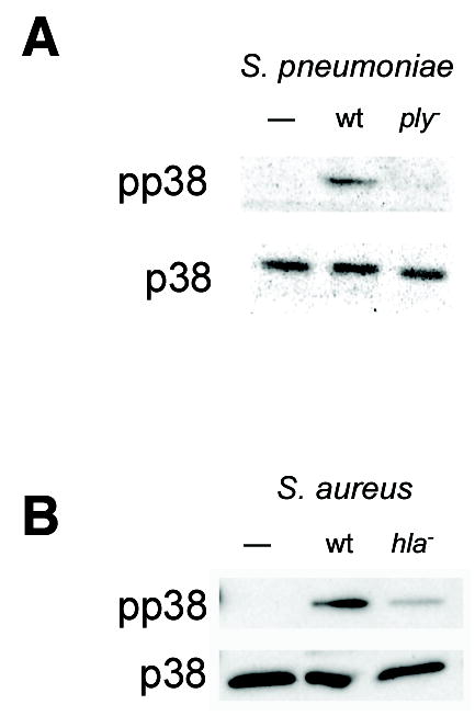
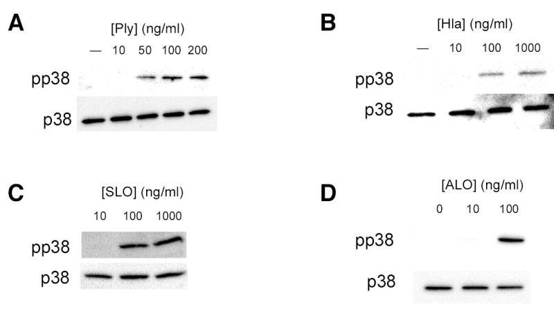
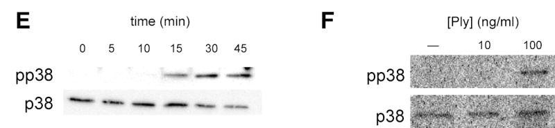
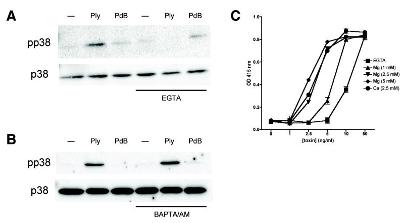
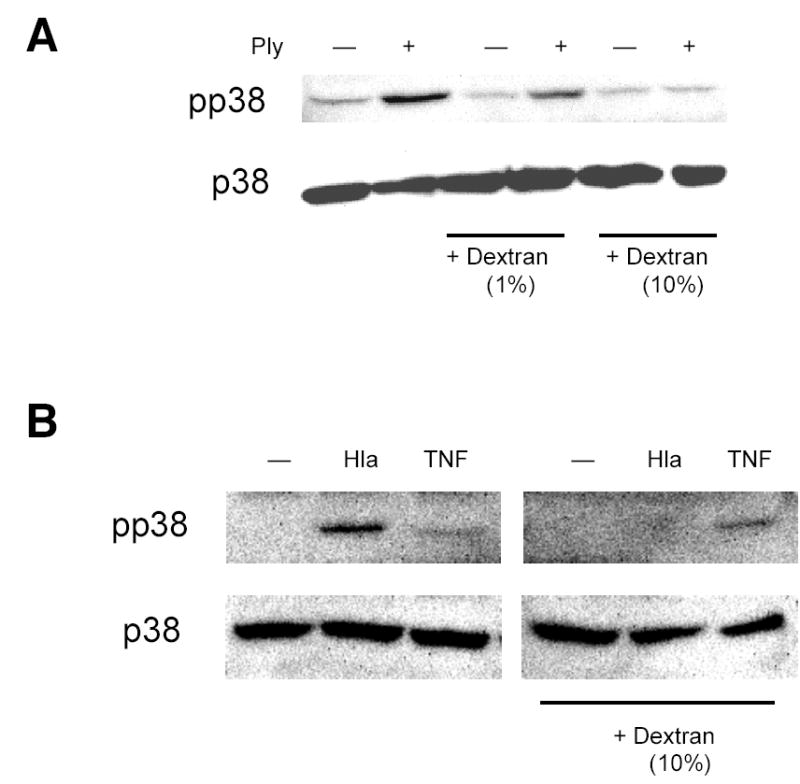
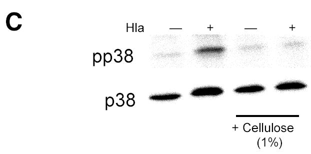
Epithelial cells act as an interface between human mucosal surfaces and the surrounding environment. As a result, they are responsible for the initiation of local immune responses, which may be crucial for prevention of invasive infection. Here we show that epithelial cells detect the presence of bacterial pore-forming toxins (including pneumolysin from Streptococcus pneumoniae, alpha-hemolysin from Staphylococcus aureus, streptolysin O from Streptococcus pyogenes, and anthrolysin O from Bacillus anthracis) at nanomolar concentrations, far below those required to cause cytolysis. Phosphorylation of p38 MAPK appears to be a conserved response of epithelial cells to subcytolytic concentrations of bacterial poreforming toxins, and this activity is inhibited by the addition of high molecular weight osmolytes to the extracellular medium. By sensing osmotic stress caused by the insertion of a sublethal number of pores into their membranes, epithelial cells may act as an early warning system to commence an immune response, while the local density of toxin-producing bacteria remains low. Osmosensing may thus represent a novel innate immune response to a common bacterial virulence strategy.


Figures

Figure 1
Figure 1
Pore-forming toxins are essential for bacterial activation of epithelial p38 MAPK. Confluent monolayers of A549 cells were stimulated for 30 min with 1×10⁷ cfu/ml of S. pneumoniae D39 or its isogenic Ply-deficient mutant, D39ply (A) or with S. aureus RN6390 or its isogenic α-hemolysin mutant, RN6390hla (B). In both cases, the wild-type, but not the toxin-deficient mutant, led to phosphorylation of p38 MAPK as assessed by Western blotting with antibodies specific for total p38 MAPK (p38) and phosphorylated p38 (pp38).
Figure 2
Figure 2
Purified bacterial pore-forming toxins activate epithelial p38 MAPK. Confluent monolayers of A549 cells were treated with the indicated concentrations of purified pneumolysin (Ply) (A), S. aureus α-hemolysin (hla) (B), streptolysin O (SLO) (C), or anthrolysin O (ALO) (D) for 30 min or with 100 ng/ml Ply for the indicated times (E). Total and phosphorylated p38 MAPK were assessed by Western blotting. Similar responses were noted in Detroit 562 nasopharyngeal epithelial cells stimulated with Ply (F).
Figure 2
Figure 2
Purified bacterial pore-forming toxins activate epithelial p38 MAPK. Confluent monolayers of A549 cells were treated with the indicated concentrations of purified pneumolysin (Ply) (A), S. aureus α-hemolysin (hla) (B), streptolysin O (SLO) (C), or anthrolysin O (ALO) (D) for 30 min or with 100 ng/ml Ply for the indicated times (E). Total and phosphorylated p38 MAPK were assessed by Western blotting. Similar responses were noted in Detroit 562 nasopharyngeal epithelial cells stimulated with Ply (F).
Figure 3
Figure 3
Pore formation is essential for Ply-mediated p38 MAPK activation and interleukin-8 release from epithelial cells. Horse erythrocytes were incubated with the indicated concentrations of purified Ply for 30 min. Optical density at 415 nm was used to assess hemoglobin release from lysed cells (A). Native Ply (100 ng/ml), but not its non-cytolytic toxoid PdB (100 ng/ml) or treatment with media alone, activates p38 MAPK in A549 cells (B). Treatment of A549 cells with Ply (100 ng/ml) but not PdB (100 ng/ml) leads to interleukin-8 release as measured by ELISA. Pretreatment of cells with SB203580, an inhibitor of p38 MAPK, abolishes this effect (C). **, P < 0.001; NS, not significant (P > 0.05).
Figure 4
Figure 4
Extracellular divalent cations are required for p38 MAPK activation and pore formation by Ply. Pretreatment of A549 cells with the extracellular divalent cation chelator EGTA (5 mM) inhibits p38 MAPK phosphorylation in response to Ply (100 ng/ml, 30 min) (A). In contrast, the cell-permeant chelator BAPTA/AM (10 μM) has no effect on toxin-mediated p38 MAPK phosphorylation (B). Horse erythrocyte lysis by Ply at the concentrations indicated is inhibited in the presence of EGTA and is restored by the addition of MgCl2 or CaCl2 to the extracellular medium (C).
Figure 5
Figure 5
Sensation of osmotic stress mediates toxin-induced p38 MAPK phosphorylation. Addition of high molecular weight dextran (M.W. 167,000) to the extracellular medium inhibits p38 MAPK phosphorylation following treatment with Ply (100 ng/ml, 30 min) (A) or hla (100 ng/ml, 30 min) (B) but not with TNF-α (10 ng/ml, 5 min) (B). Cellulose suspension (20 μm particle size, 1%) inhibits p38 MAPK phosphorylation in A549 cells exposed to hla (100 ng/ml, 30 min).
Figure 5
Figure 5
Sensation of osmotic stress mediates toxin-induced p38 MAPK phosphorylation. Addition of high molecular weight dextran (M.W. 167,000) to the extracellular medium inhibits p38 MAPK phosphorylation following treatment with Ply (100 ng/ml, 30 min) (A) or hla (100 ng/ml, 30 min) (B) but not with TNF-α (10 ng/ml, 5 min) (B). Cellulose suspension (20 μm particle size, 1%) inhibits p38 MAPK phosphorylation in A549 cells exposed to hla (100 ng/ml, 30 min).

References

    1. Burns DL. ASM Press; Washington, D.C: 2003. Bacterial protein toxins.
    1. Braun JS, Sublett JE, Freyer D, Mitchell TJ, Cleveland JL, Tuomanen EI, Weber JR. J Clin Invest. 2002;109(1):19–27. - PMC - PubMed
    1. Berry AM, Yother J, Briles DE, Hansman D, Paton JC. Infect Immun. 1989;57(7):2037–2042. - PMC - PubMed
    1. Kernodle DS, Voladri RK, Menzies BE, Hager CC, Edwards KM. Infect Immun. 1997;65(1):179–184. - PMC - PubMed
    1. Limbago B, Penumalli V, Weinrick B, Scott JR. Infect Immun. 2000;68(11):6384–6390. - PMC - PubMed

Publication types

MeSH terms