. 2006 Apr;9(4):511-8.
doi: 10.1038/nn1666. Epub 2006 Mar 5.

UNC-6/Netrin induces neuronal asymmetry and defines the site of axon formation

Affiliations

UNC-6/Netrin induces neuronal asymmetry and defines the site of axon formation

Carolyn E Adler et al. Nat Neurosci. 2006 Apr.

Abstract

UNC-6/Netrin and its receptor UNC-40/DCC are conserved regulators of growth cone guidance. By directly observing developing neurons in vivo, we show that UNC-6 and UNC-40 also function during axon formation to initiate, maintain and orient asymmetric neuronal growth. The immature HSN neuron of Caenorhabditis elegans breaks spherical symmetry to extend a leading edge toward ventral UNC-6. In unc-6 and unc-40 mutants, leading edge formation fails, the cell remains symmetrical until late in development and the axon that eventually forms is misguided. Thus netrin has two activities: one that breaks neuronal symmetry and one that guides the future axon. As the axon forms, UNC-6, UNC-40 and the lipid modulators AGE-1/phosphoinositide 3-kinase (PI3K) and DAF-18/PTEN drive the actin-regulatory pleckstrin homology (PH) domain protein MIG-10/lamellipodin ventrally in HSN to promote asymmetric growth. The coupling of a directional netrin cue to sustained asymmetric growth via PI3K signaling is reminiscent of polarization in chemotaxing cells.


Figures

Figure 1
Figure 1
Development of HSN in wild-type C. elegans. (a) HSN expresses UNC-40/DCC receptors, which guide the axon towards UNC-6/netrin in the ventral nerve cord (VNC). (b) Mature HSN, late L4 stage. Axon extends ventrally, then anteriorly, and defasciculates from the VNC to form synapses at the vulva, (c–i) Lateral views of HSN in wild-type larvae expressing unc-86::myr-GFP, paired with schematic diagrams. Green, HSN; gray, PLM axon, which also expresses unc-86::myr-GFP. Hours of development and developmental stage are noted, (c) Early L1. HSN extends filopodia in random directions (arrows), (d) Early L2. HSN is polarized ventrally. (e) Late L2. Ventral side of HSN has a dynamic leading edge, (f) Early L3. HSN cell body has migrated ventrally, leaving a dorsal retraction fiber (arrowhead), (g) Mid-L3. HSN extends multiple neurites (arrows) toward the VNC (arrowhead), (h) L3-L4 transition. One growth cone expands in size and the others retract, (i) Mid-L4. The growth cone has turned anteriorly after reaching the VNC. In all pictures, ventral is down and anterior is to the left. Scale bars: 10 μm in b; 5 μm in c–i.
Figure 2
Figure 2
HSN neurites are microtubule-containing, actin-rich structures. (a,b) HSN reconstructions based on serial EM sections, (a) Mid-L3 HSN with multiple neurites. (b) L3-L4 transition HSN with a single axon. Anterior is to the left and ventral is down, (c–f) Electron micrographs of HSN neurites. The position and orientation of the micrographs are indicated by the labeled vertical lines in a and b. In each section, a red bar marks the position of the HSN neurite. From left to right, each section includes muscle, basement membrane, the HSN neurite, epidermis and cuticle. Medial is to the left and ventral is down. In c and d, cross-sections of a neurite from a. (c) Proximal region showing microtubules in tangential view (arrows). Arrowheads span a stretch of basement membrane, (d) Distal region enriched in filaments (arrowheads). In e and f, cross-sections of a neurite from b. (e) Proximal region containing microtubules in tangential view (arrows) and ribosomes (fat arrows), (f) Distal region enriched in filaments (arrowheads) and ribosomes (fat arrows). In g and h, HSN neurons expressing GFP-actin at mid-L3 (g) and L3–L4 transition (h). Fluorescence is enriched in the distal regions of the neurites (arrowheads). Anterior is to the left and ventral is down. Scale bars: 200 nm in c–f; 5 μm in g,h.
Figure 3
Figure 3
HSN development in netrin mutants. HSN development in wild-type, unc-6(ev400) and unc-40(e271) C. elegans. HSN was visualized with unc-86::myr-GFP. (a–c) Percentage of HSNs with filopodia or leading edges in the L1 and L2 stages. An HSN was scored as polarized if it had any filopodia or a leading edge, and the most prevalent direction of growth was noted. HSNs in unc-6 and unc-40 mutants had filopodia at early time points but did not form a leading edge at any time during this interval. (d–f) L2 stage HSNs. (g–i) L3 stage HSNs. (j) Percentage of HSN neurons with predominantly ventral neurites in mid-L3. (k–m) L4 stage HSNs. (n) Percentage of HSN neurons with a single axon in mid-L4. Error bars represent the standard error of proportion. For panels a–c, j and n, n ≥ 100 cells. All images are lateral views with ventral down and anterior to the left. Scale bars, 5μm.
Figure 4
Figure 4
Heat-inducible expression of UNC-6/netrin induces leading edge formation in HSN. (a) Immunoblot with anti-HA antibody of unc-6(ev400) mutants expressing hs::unc-6::HA, raised at 15 °C (−) or exposed to heat shock at 30 °C for 2 h (+). (b,c) HSN in wild-type L4 larvae expressing hs::unc-6::HA. (b) Control L4 larva raised at 15 °C. (c) Heat shock causes axon wandering. (d,e) Gonads of wild-type L4 larvae expressing hs::unc-6::HA. (d) Control L4 larva raised at 15 °C. (e) Exposure to heat shock causes abnormal gonad morphology, marked by clear area (arrows). Asterisks indicate the developing vulva. (f,g) HSN in early L2 unc-6 mutants expressing hs::unc-6::HA. (f) Control L2 larva raised at 15 °C, with unpolarized HSN morphology. (g) L2 larva exposed to 2 h heat shock during late L1 and examined in early L2. HSN has a polarized leading edge oriented in the wrong (posterior) direction. (h) Percentage of polarized HSNs after heat shock. A cell was scored as polarized if it had one leading edge (as in g) or if it had multiple filopodia oriented in one direction. ***P < 0.001 as determined by the t-test for proportions. For each condition, n ≥ 100 cells. Error bars represent standard error of proportion. The polarization defect in unc-6 and unc-40 mutants was less severe here than in Figure 3, probably because the larvae were raised at 15 °C instead of 25 °C. In all pictures, anterior is to the left and ventral is down. Scale bars: 10 μm in b and c; 5 μm in f and g.
Figure 5
Figure 5
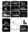
UNC-40::GFP and MIG-10::YFP are ventrally localized in HSN. (a–c) myr-GFP is evenly distributed in HSN membranes in the L1 and L2 stages, (d–f) UNC-40::GFP is preferentially localized to the ventral HSN in the L2 stage, (g–i) MIG-10::YFP is preferentially localized to the ventral HSN in the late L2 stage, (j–k) Lateral views of L2 stage HSNs expressing UNC-40::GFP. (j) unc-40 mutant rescued by the unc-40::GFP transgene. (k) unc-6 mutant. Traces are line-scan intensity plots (in arbitrary units) of the GFP signal around the periphery of the cell shown, starting from the arrow and moving clockwise. (l) Average ratio of dorsal to ventral intensity for > 10 cells of each genotype, unc-6 was statistically different from the control strain, a unc-40 mutant rescued with the unc-40::GFP transgene (P < 0.05). Wild-type nematodes gave qualitatively similar results to rescued unc-40 but were not quantified. Error bars indicate s.e.m. (m) UNC-40::GFP in mig-10 mutant, same stage as in panels j and k. In all pictures, ventral is down and anterior is to the left. Scale bars, 5μm.
Figure 6
Figure 6
MIG-10::YFP localizes ventrally in a netrin-dependent manner.(ag) Lateral views of L3 stage larvae expressing unc-86::YFP::mig-10. The HSN cell body fills most of each panel. Traces are line-scan intensity plots (in arbitrary units) of the YFP signal around the periphery of the cell shown, beginning at the arrow and moving clockwise. IR, intensity ratio. In b, mig-10 mutant rescued by the mig-10::YFP transgene. (h) Average intensity ratios of MIG-10::YFP signals. Ratio was calculated as the intensity of the ventral half of the perimeter divided by the dorsal half (n = 9–14 cells for each genotype). The random distribution of myr-GFP in HSN is included as a control. Error bars indicate s.e.m. By Bonferroni t-tests (P < 0.05), wild type, mig-10 and unc-34 were indistinguishable; unc-6, unc-40and daf-18 were highly defective; and age-1 was intermediate. In all pictures, ventral is down and anterior is to the left. Scale bar, 5 μm.

References

    1. Dotti CG, Sullivan CA, Banker GA. The establishment of polarity by hippocampal neurons in culture. J Neurosci. 1988;8:1454–1468. - PMC - PubMed
    1. Inagaki N, et al. CRMP-2 induces axons in cultured hippocampal neurons. Nat Neurosci. 2001;4:781–782. - PubMed
    1. Shi SH, Cheng T, Jan LY, Jan YN. APC and GSK-3β are involved in mPar3 targeting to the nascent axon and establishment of neuronal polarity. Curr Biol. 2004;14:2025–2032. - PubMed
    1. Kishi M, Pan YA, Crump JG, Sanes JR. Mammalian SAD kinases are required for neuronal polarization. Science. 2005;307:929–932. - PubMed
    1. Shi SH, Jan LY, Jan YN. Hippocampal neuronal polarity specified by spatially localized mPar3/mPar6 and PI 3-kinase activity. Cell. 2003;112:63–75. - PubMed

Publication types

MeSH terms