. 2006 Feb 14;12(6):928-34.
doi: 10.3748/wjg.v12.i6.928.

Cyclooxygenase-2 and epithelial growth factor receptor up-regulation during progression of Barrett's esophagus to adenocarcinoma

Affiliations

Cyclooxygenase-2 and epithelial growth factor receptor up-regulation during progression of Barrett's esophagus to adenocarcinoma

Yan Li et al. World J Gastroenterol. .

Abstract

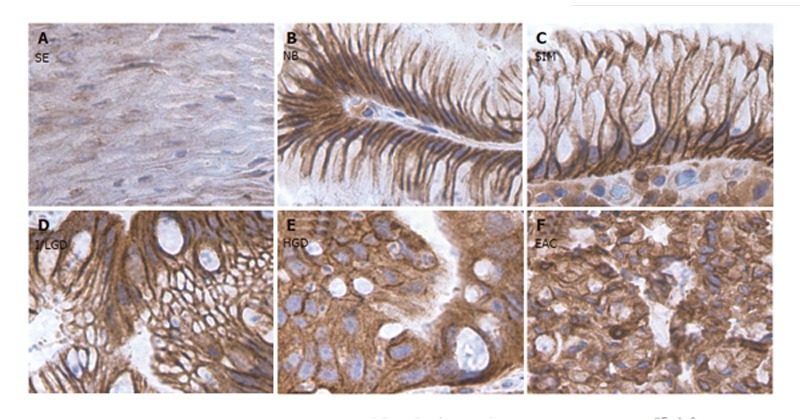
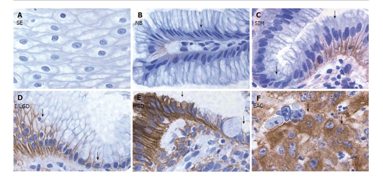
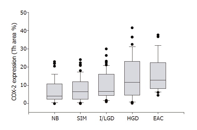
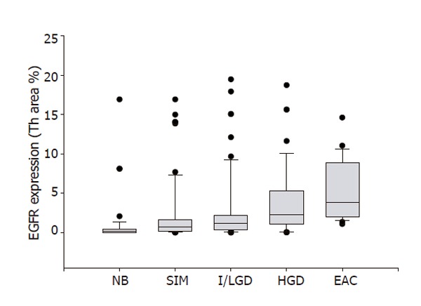
Aim: To investigate the expression of cyclooxygenase-2 (COX-2) and epithelial growth factor receptor (EGFR) throughout the progression of Barretts esophagus (BE).

Methods: COX-2 and EGFR protein expressions were detected by using immunohistochemical method. A detailed cytomorphological changes were determined. Areas of COX-2 and EGFR expression were quantified by using computer Imaging System.

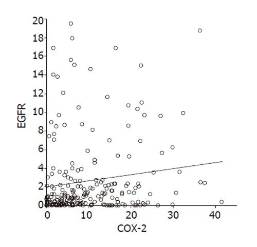
Results: The expressions of both COX-2 and EGFR increased along with the progression from BE to esophagus adenocarcinoma (EAC). A positive correlation was found between COX-2 expression and EGFR expression.

Conclusion: COX-2 and EGFR may be cooperative in the stepwise progression from BE to EAC, thereby leading to carcinogenesis.


Figures

Figure 1
Figure 1
COX-2 staining in the sections from different stages of BE along with the sections from normal squamous and CLE nondetectable BE. A: Squamous epithelium (SE); B: Nondetectable BE in CLE (NB); C: Specialized intestinal metaplasia (SIM); D: Indeterminate/low-grade dysplasia (I/LGD); E: high-grade dysplasia (HGD); F: Esophageal adenocarcinoma (EAC). Strong COX-2 staining was detected in both NCP cells and ACP cells in the histological stages of SIM, I/LGD, HGD and EAC. A weak COX-2 staining was observed in the normal squamous epithelium.
Figure 2
Figure 2
EGFR staining in the sections from different stages of BE along with the sections from normal squamous and CLE nondetectable BE. A: Squamous epithelium (SE); B: Nondetectable BE in CLE (NB); C: Specialized intestinal metaplasia (SIM); D: Indeterminate/low-grade dysplasia (I/LGD); E: high-grade dysplasia (HGD); F: Esophageal adenocarcinoma (EAC). Strong EGFR staining was detected in ACP cells (arrowhead) in the histological stages of SIM, I/LGD, HGD and EAC. A weak EGFR staining was found in NCP cells (arrow) and in the normal squamous epithelium.
Figure 3
Figure 3
COX-2 expression and pathological severity of BE. COX-2 expression is plotted against the 4 stages of Barrett's esophagus. The line enclosed within the box represents the median value; the small circles indicate outlying data points. COX-2 expression in specimens with SIM, I/LGD, HGD and EAC was significantly increased in comparison with that in NB (P < 0.01). A significant positive correlation between COX-2 expression and histopathologic changes from SIM to EAC was observed (r = 0.97, P < 0.001).
Figure 4
Figure 4
EGFR expression and pathological severity of BE. EGFR expression is plotted against the 4 stages of Barrett's esophagus. The line enclosed within the box represents the median value; the small circles indicate outlying data points. EGFR expression in specimens with SIM, I/LGD, HGD and EAC was significantly increased in comparison with that in NB (P < 0.01). A significant positive correlation between EGFR expression and histopathologic changes from SIM to EAC was found(r = 0.97, P < 0.001).
Figure 5
Figure 5
Relationship between COX-2 expression and EGFR expression in 104 patients. A significant positive correlation between COX-2 expression and EGFR expression was found (r = 0.94, P < 0.001).

References

    1. Blot WJ, Devesa SS, Kneller RW, Fraumeni JF Jr. Rising incidence of adenocarcinoma of the esophagus and gastric cardia. JAMA. 1991;265:1287–1289. - PubMed
    1. Pera M, Cameron AJ, Trastek VF, Carpenter HA, Zinsmeister AR. Increasing incidence of adenocarcinoma of the esophagus and esophagogastric junction. Gastroenterology. 1993;104:510–513. - PubMed
    1. Farrow DC, Vaughan TL. Determinants of survival following the diagnosis of esophageal adenocarcinoma (United States) Cancer Causes Control. 1996;7:322–327. - PubMed
    1. Isolauri J, Luostarinen M, Isolauri E, Reinikainen P, Viljakka M, Keyriläinen O. Natural course of gastroesophageal reflux disease: 17-22 year follow-up of 60 patients. Am J Gastroenterol. 1997;92:37–41. - PubMed
    1. Altorki NK, Oliveria S, Schrump DS. Epidemiology and molecular biology of Barrett's adenocarcinoma. Semin Surg Oncol. 1997;13:270–280. - PubMed

Publication types

MeSH terms