Review
. 2006;7(2):206.
doi: 10.1186/gb-2006-7-2-206. Epub 2006 Feb 28.

The aquaporins

Affiliations
Review

The aquaporins

Elisabeth Kruse et al. Genome Biol. 2006.

Abstract

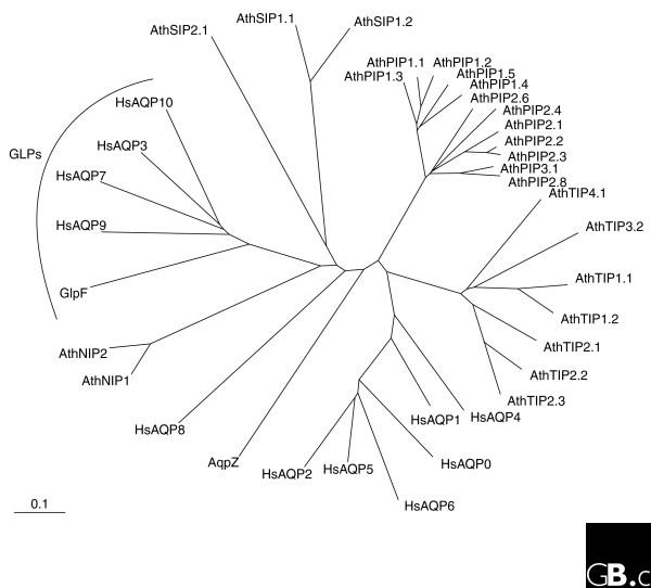
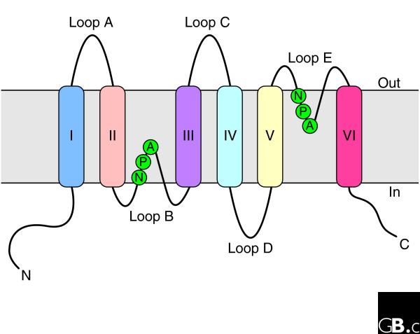
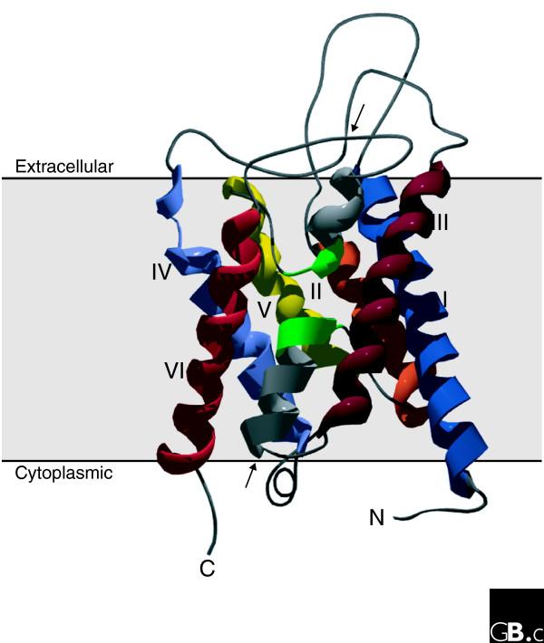
Water is the major component of all living cells, and efficient regulation of water homeostasis is essential for many biological processes. The mechanism by which water passes through biological membranes was a matter of debate until the discovery of the aquaporin water channels. Aquaporins are intrinsic membrane proteins characterized by six transmembrane helices that selectively allow water or other small uncharged molecules to pass along the osmotic gradient. In addition, recent observations show that some aquaporins also facilitate the transport of volatile substances, such as carbon dioxide (CO2) and ammonia (NH3), across membranes. Aquaporins usually form tetramers, with each monomer defining a single pore. Aquaporin-related proteins are found in all organisms, from archaea to mammals. In both uni- and multicellular organisms, numerous isoforms have been identified that are differentially expressed and modified by post-translational processes, thus allowing fine-tuned tissue-specific osmoregulation. In mammals, aquaporins are involved in multiple physiological processes, including kidney and salivary gland function. They are associated with several clinical disorders, such as kidney dysfunction, loss of vision and brain edema.


Figures

Figure 1
Figure 1
The evolutionary relationships of aquaporins. A phylogenetic tree was generated from human (Hs), Arabidopsis (Ath) and E. coli (AqpZ and GlpF) aquaporin sequences using ClustalX. Members of the aquaglyceroporin (GLP) subfamily are indicated; all other proteins shown belong to the classical aquaporin subfamily.
Figure 2
Figure 2
Topology of an aquaporin protein within the membrane. The protein consists of six transmembrane helices (I-VI) connected by five loops (A-E) and includes two internal tandem repeats (I-III and IV-VI, respectively). Loops B and E, containing the conserved NPA motifs (in the single-letter amino-acid code), form short α helices that fold back into the membrane from opposite sides. C, carboxyl terminus; N, amino terminus.
Figure 3
Figure 3
Three-dimensional structure of an aquaporin subunit monomer (a ribbon model of NtAQP1, a PIP1 protein from tobacco). The structure shows six tilted membrane-spanning helices (I-VI) and two pore-forming domains made up of two short α helices entering the membrane from the extracellular and intracellular surfaces (arrows). The two NPA boxes are indicated in green. Amino- and carboxy-terminal domains are oriented to the cytoplasmic side of the membrane. The figure was generated using MODELLER7v7 and Swiss-Pdb Viewer.

References

    1. Heymann JB, Engel A. Aquaporins: phylogeny, structure, and physiology of water channels. News Physiol Sci. 1999;14:187–193. - PubMed
    1. Hohmann S, Bill RM, Kayingo G, Prior BA. Microbial MIP channels. Trends Microbiol. 2000;8:33–38. doi: 10.1016/S0966-842X(99)01645-5. - DOI - PubMed
    1. Murata K, Mitsuoka K, Hirai T, Walz T, Agre P, Heymann JB, Engel A, Fujiyoshi Y. Structural determinants of water permeation through aquaporin-1. Nature. 2000;407:599–605. doi: 10.1038/35036519. - DOI - PubMed
    1. Fu DX, Libson A, Miercke LJW, Weitzman C, Nollert P, Krucinski J, Stroud RM. Structure of a glycerol-conducting channel and the basis for its selectivity. Science. 2000;290:481–486. doi: 10.1126/science.290.5491.481. - DOI - PubMed
    1. Zardoya R. Phylogeny and evolution of the major intrinsic protein family. Biol Cell. 2005;97:397–414. - PubMed

LinkOut - more resources