. 2006 Mar 6;34(5):e37.
doi: 10.1093/nar/gkl034.

Control of small inhibitory RNA levels and RNA interference by doxycycline induced activation of a minimal RNA polymerase III promoter

Affiliations

Control of small inhibitory RNA levels and RNA interference by doxycycline induced activation of a minimal RNA polymerase III promoter

Lahouari Amar et al. Nucleic Acids Res. .

Abstract

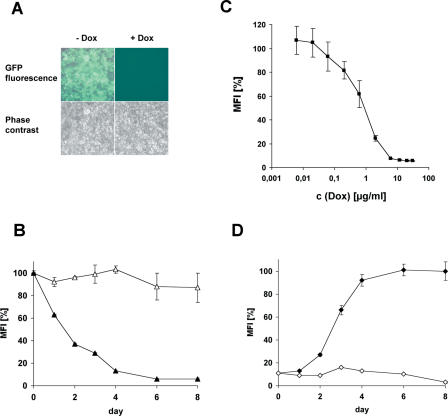
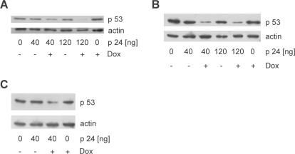
RNA interference (RNAi) mediated by expression of short hairpin RNAs (shRNAs) is a powerful tool for efficiently suppressing target genes. The approach allows studies of the function of individual genes and may also be applied to human therapy. However, in many instances regulation of RNAi by administration of a small inducer molecule will be required. To date, the development of appropriate regulatory systems has been hampered by the few possibilities for modification within RNA polymerase III promoters capable of driving efficient expression of shRNAs. We have developed an inducible minimal RNA polymerase III promoter that is activated by a novel recombinant transactivator in the presence of doxycycline (Dox). The recombinant transactivator and the engineered promoter together form a system permitting regulation of RNAi by Dox-induced expression of shRNAs. Regulated RNAi was mediated by one single lentiviral vector, blocked the expression of green fluorescent protein (GFP) in a GFP-expressing HEK 293T derived cell line and suppressed endogenous p53 in wild-type HEK 293T, MCF-7 and A549 cells. RNA interference was induced in a dose- and time-dependent manner by administration of Dox, silenced the expression of both target genes by 90% and was in particular reversible after withdrawal of Dox.


Figures

Figure 1
Figure 1
Schematic diagrams illustrating the regulatory system allowing Dox-induced RNAi. (A) Primary structure of the transactivator rtTA-Oct2 composed of the conditional DNA binding domain of rtTA2-M2, and the Oct-2Q(Q→A) domain mediating specific induction of a minimal RNA polymerase III promoter (17). (B) Structure of the minimal U6 promoter: The 202 bp sequence upstream from the transcription start site was derived from the human U6 promoter and contains the PSE and the TATA box. Upstream from this sequence, seven tet operator sequences (tetOs) have been inserted to allow conditional binding of the transactivator. (C) In the absence of Dox (off state), rtTA-Oct2 does not bind to the operator sequences and hence shRNAs are not synthesized. (D) In the presence of Dox (on state), the transactivator binds and thereby activates the expression of shRNAs designed to induce the degradation of the respective target mRNAs.
Figure 2
Figure 2
A single lentiviral vector mediates Dox-regulated RNAi. (A) Design of the vector: LTR, ψ and Flap are sequences derived from HIV-1 (the LTRs, the packaging sequence and the central Flap element, respectively). P U6 min and P PGK are the Tet-regulated minimal U6 promoter and the phosphoglycerate kinase promoter; WPRE is the Woodchuck hepatitis virus responsive element; rtTA-Oct2, the cDNA encoding the transcription factor rTA-Oct2; and shRNA, the sequence encoding shRNAs. (B and C) Experimental validation of regulated RNAi using a vector that expresses shRNAs designed to silence the expression of GFP. (B) ‘Northern blot’ analysis of Dox-regulated expression of siRNAs from the vector. HEK 293T GFP cells (1 × 10⁵) were incubated for 24 h with and without vector corresponding to 141 ng of protein p24, and cultivated in the presence and absence of 6 µg/ml Dox for 7 days. Then, small RNAs were isolated from the cells and probed for siRNAs designed to silence the expression of GFP. 5S-rRNA detected by ethidium bromide staining of the polyacrylamide gel served as an internal control to show equal loading. (C) Experimental validation of RNAi-mediated silencing of GFP. HEK 293T GFP cells (8 × 10⁴) were incubated overnight with various quantities of vector expressed as ng of protein p24, and cultivated in the absence (grey bars) and in the presence (white bars) of 6 µg/ml Dox for 5 days prior to FACS analysis. Values are averages of percentages of GFP-positive cells ± SE, n = 3.
Figure 3
Figure 3
Characterization of Dox-regulated RNAi in a representative cell-clone (C9): (A) Microscopic analysis of cells incubated in the presence or in the absence of 6 µg/ml Dox at 72 h after induction. (B) Time course of Dox-induced RNAi: RNAi was induced or not induced at day 0 by administration of 6 µg/ml Dox and mean intensities of GFP fluorescence were measured by FACS analysis at various times after induction. Closed triangles represent intensities of cells incubated with Dox, open triangles give those of untreated cells. The fluorescence intensity observed at day 0 was defined as 100%, values are means ± SE, n = 3. (C) Mean intensities (± SE, n = 3) of GFP fluorescence obtained by FACS analysis of cells cultivated for 5 days in the presence of various concentrations of Dox. The fluorescence intensity in untreated cells was defined as 100%. (D) Reappearance of GFP fluorescence after withdrawal of Dox: prior to the analysis, cells were cultivated for 5 days in the presence of 6 µg/ml Dox. At day 0, Dox was withdrawn or not withdrawn and the mean fluorescence intensity was followed by FACS analysis. Closed rhomboids represent values from cells that were not treated with Dox from day 0, open rhomboids give values from cells incubated with 6 µg/ml Dox throughout the experiment. The fluorescence intensity measured 8 days after removal of Dox was defined as 100%, values are means ± SE, n = 3.
Figure 4
Figure 4
‘Western blot’ analysis demonstrating silencing of p53 by Dox-regulated RNAi in (A) HEK 293T cells, (B) MCF-7 cells and (C) A549 cells. Cells (1 × 10⁵) were incubated overnight with indicated quantities of vector, expressed as ng of protein p24, and then cultivated in the absence and in the presence of 6 µg/ml Dox. After a 5 day (MCF-7 and A549 cells) and a 7 day cultivation (HEK 293T cells), protein was extracted from the cells and analyzed by immunoblotting. Both p53 and actin were detected; the latter served as a control to demonstrate equal loading.

References

    1. Hannon G.J. RNA interference. Nature. 2002;418:244–251. - PubMed
    1. Brummelkamp T.R., Bernards R., Agami R. A system for stable expression of short interfering RNAs in mammalian cells. Science. 2002;296:550–553. - PubMed
    1. Xia H., Mao Q., Paulson H.L., Davidson B.L. siRNA mediated gene silencing in vitro and in vivo. Nat. Biotechnol. 2002;20:1006–1010. - PubMed
    1. Abbas-Terki T., Blanco-Bose W., Déglon N., Pralong W., Aebischer P. Lentiviral-mediated RNA interference. Hum. Gene Ther. 2002;13:2197–2201. - PubMed
    1. Tiscornia G., Singer O., Ikawa M., Verma I.M. A general method for gene knockdown in mice by using lentiviral vectors expressing small interfering RNA. Proc. Natl Acad. Sci. USA. 2003;100:1844–1848. - PMC - PubMed

Publication types