. 2006 Mar 6;34(5):1405-15.
doi: 10.1093/nar/gkl032. Print 2006.

Human replication protein A can suppress the intrinsic in vitro mutator phenotype of human DNA polymerase lambda

Affiliations

Human replication protein A can suppress the intrinsic in vitro mutator phenotype of human DNA polymerase lambda

Giovanni Maga et al. Nucleic Acids Res. .

Abstract

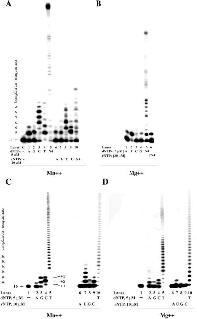
DNA polymerase lambda (pol lambda) is a member of the X family DNA polymerases and is endowed with multiple enzymatic activities. In this work we investigated the in vitro miscoding properties of full-length, human pol lambda either in the absence or in the presence of the human auxiliary proteins proliferating cell nuclear antigen (PCNA) and replication protein A (RP-A). Our data suggested that (i) pol lambda had an intrinsic ability to create mismatches and to incorporate ribonucleotides at nearly physiological Mn++ and Mg++ concentrations; (ii) the sequence of the template-primer could influence the misincorporation frequency of pol lambda; (iii) pol lambda preferentially generated G:T and G:G mismatches; (iv) RP-A, but not PCNA, selectively prevented misincorporation of an incorrect nucleotide by pol lambda, without affecting correct incorporation and (v) this inhibitory effect required a precise ratio between the concentrations of pol lambda and RP-A. Possible physiological implications of these findings for the in vivo fidelity of pol lambda are discussed.


Figures

Figure 1
Figure 1
Pol λ can misincorporate deoxy- and ribonucleotides in the presence of physiological Mn++ and Mg++ concentrations. The sequences of the primer/templates used are indicated in Materials and Methods. (A) Reactions were performed as described in Materials and Methods, in the presence of 50 nM (0.5 pmol) pol λ, 0.1 mM Mn++ and each of the four dNTPs at 5 µM final concentration. Lanes 1–4: 5′-labelled 18/40merA primer/template; lanes 6–9: 19/40merT; lanes 11–14: 20/40merG; lanes 16–19: 21/40merC. Lanes 5, 10, 15 and 20: control reactions with all four dNTPs at 5 µM each. Lanes 21–24: control reactions in the absence of dNTPs. Asterisks indicate the mismatches. (B) The experiment was performed as in panel A, but in the presence of 1 mM Mg++. The migration of four different primers is shown on the right side of the panel. Asterisks indicate the mismatches. (C) Reactions were performed as described in Materials and Methods in the presence of 50 nM (0.5 pmol) pol λ, 0.1 mM Mn++ (lanes 1–20) or 1 mM Mg++ (lanes 21–39) and each of the four rNTPs at a final concentration of 10 µM. Lanes 1–4; 21–24: 5′-labelled 18/40merA primer/template; lanes 17–20; 25–28: 19/40merT; lanes 11–15, 30–33: 20/40merG; lanes 16–19; 36–39: 21/40merC. Lanes 9, 15 and 34: reactions in the presence of a mixture of all four rNTPs at 10 µM each. Lanes 10, 16, 29 and 35: control reactions in the presence of all four dNTPs at 5 µM each final concentration. The running position of the four different primers is indicated on the left side of the panel.
Figure 2
Figure 2
The sequence context influences misincorporation by pol λ. All reactions were performed as described in Materials and Methods in the presence of 50 nM (0.5 pmol) pol λ. (A) Incorporation of the four dNTPs (lanes 1–4, 5 µM final concentration) or the four rNTPs (lanes 6–9, 20 µM final concentration) on the 5′-labelled 17/75mer primer template, in the presence of 0.1 mM Mn++. Lane C: control reaction in the absence of dNTPs or rNTPs. Lane 5: reaction in the presence of all four dNTPs (5 µM each). Lane 10: reaction in the presence of all four rNTPs (20 µM each). The position of the 17mer primer and the sequence of the first six positions on the template strand are indicated on the left side of the panel. (B) Reaction conditions were the same as for panel A, but 1 mM Mg++ was used instead of Mn++. The four dNTPs were added individually (lanes 1–4) or all together (lane 5) at a final concentration of 5 µM each. Lane 6: reaction in the presence of all four rNTPs at 20 µM final concentration each. (C) Reaction conditions were as in panel A, but the 5′-labelled d(T)16/d(A)200 primer/template was used. All four dNTPs (lanes 2–5) or rNTPs (lanes 6–9) were tested separately at a final concentration of 5 µM for dNTPs and 10 µM for rNTPs, in the presence of 0.1 mM Mn++. (D) Reaction conditions were the same as C, but 1 mM Mg ++ was used instead of Mn++.
Figure 3
Figure 3
Pol λ appears to preferentially generate G:T over C:T mismatches and its misincorporation ability is suppressed by RP-A. (A) Misincorporation of dGTP (lanes 1–3) or dCTP (lanes 4–6) was monitored on the 19/40merT template in the presence of 0.1 mM Mn++ and increasing concentrations of pol λ (25, 100 and 250, corresponding to 0.25, 1 and 2.5 pmol, respectively). Lane 7: control reaction in the presence of 25 nM pol λ and all four dNTPs at 5 µM each. The positions of the differently migrating dGMP- and dCMP-terminated +1 products are shown on the left side of the panel. (B) Misincorporation of increasing concentrations of dGTP (lanes 1–6) or dCTP (lanes 7–12) were monitored on the 19/40merT template in the presence of 0.1 mM Mn++ and 25 nM (0.25 pmol) pol λ. The positions of the differently migrating dGMP- and dCMP-terminated +1 products are shown on the left side of the panel. (C) Incorporation of dTTP or dATP (5 µM each) was measured in the presence of 50 nM (0.5 pmol) polλ, 5′-labelled d(T)16/d(A)200 primer/template, 1 mM Mg++ and in the absence (lanes 1 and 9) or in the presence of different concentrations of either PCNA (lanes 2, 3, 10 and 11) or RP-A (lanes 4–6, 12–14), or an equimolar combination of both proteins (lanes 7, 8, 15 and 16). (D) Incorporation of 5 µM dCTP (lanes 1–8) or 10 µM dGTP (lanes 9–16) was monitored on the 5′-labelled 20/40merG primer/template, in the presence 150 nM (1.5 pmol) pol λ, 0.1 mM Mn++, and in the absence (lanes 1 and 9) or in the presence of 0.5 (lanes 2, 10) and 2 pmol (lanes 3, 11) of PCNA, or 0.2 (lanes 4, 12), 0.5 (lanes 5, 13) and 2 pmol (lanes 6, 14) of RP-A, or a combination of 0.2 (lanes 7, 15) or 2 pmol (lanes 15, 16) of PCNA and RP-A in equimolar ratios. The positions of the differently migrating dCMP- and dGMP-terminated primers are indicated on the left side of the panel with an asterisc.
Figure 4
Figure 4
A precise balance of the relative concentrations of RP-A and pol λ is required for supression of misincorporation. All reactions were performed as described in Materials and Methods in the presence of 0.1 mM Mn++ and the 5′-labelled 19/40merT template. (A) Incorporation of 5 µM dATP (lanes 1–5, 11–15, 21–25) or 5 µM dGTP (lanes 6–10, 16–20, 26–30), was monitored in the presence of 0.2 (lanes 1–10), 0.5 (lanes 11–20) and 1.5 pmol (lanes 21–30) of pol λ, corresponding to final enzyme concentrations of 20, 50 and 150 nM, respectively. Reactions were performed in the absence (lanes 1, 6, 11, 16, 21 and 26) or in the presence (lanes 2–5, 7–10, 12–15, 17–20, 22–25, 26–30) of increasing amounts of RP-A, as indicated in the panel. The resulting molar/molar (M/M) ratios of RP-A to pol λ in the different reactions are also indicated. (B) Misincorporated dGMP, in the absence or in the presence of RP-A, was quantified from the experiment shown in panel A. The normalized values, expressed as percentages of the total dGMP incorporated in the absence of RP-A in each set of reactions, were plotted as a function of the RP-A concentrations. Three different exposition times were used to collect images representative of the entire dynamic range of the X-ray film sensitivity. Images were scanned independently and the mean values of the three independent measurements were used for the plot. Error bars indicate +/− SD; circles, values calculated in the presence of 0.2 pmol (20 nM) DNA pol λ; squares, values calculated in the presence of 0.5 pmol (50 nM) DNA pol λ; triangles, values calculated in the presence of 1.5 pmol (150 nM) pol λ. The actual misincorporation values in the absence of RP-A (expressed as percentage elongation of the input DNA substrate) were 4.5 (±0.5)% with 0.2 pmol pol λ, 15.8 (±0.8)% with 0.5 pmol DNA pol λ and 37.5 (±0.8)% with 1.5 pmol DNA pol λ. (C) As a control, incorporation of dATP (lanes 1–9) or dGTP (lanes 10–18) in the presence of 0.1 mM Mn++ and 0.2 pmol (20 nM) of pol λ was monitored in the absence (lanes 1 and 10) or in the presence of increasing amounts of PCNA (lanes 2–5, 11–14) or BSA (lanes 6–9, 15–18). Lane 19: control reaction in the absence of dNTPs.
Figure 5
Figure 5
(A)The intrinsic mutator phenotype of pol λ is not affected by the combination of Mn++ and Mg++ over a wide range of molar ratios. Incorporation of dTTP, dATP or dGTP were monitored in the presence of 100 nM d(T)16/d(A)200 and 50 nM pol λ, under the reactions conditions described in Materials and Methods. Kinetic parameters (kcat, Km, kcat/Km and 1/f) were determined as described in Materials and Methods. Nucleotide concentrations used were 0.5, 2.5, 10, 20 and 100 µM, respectively. A. Incorporation efficiencies (kcat/Km) values for dTTP (white bars), dATP (light grey bars) and dGTP (dark grey bars), were determined in the presence of 0.1 mM Mn++, either alone or in combination with 0.1, 0.5 or 2 mM Mg++ respectively, or in the presence of 2 mM Mg++ alone. (B) Fidelity values for dATP (light grey bars) and dGTP (dark grey bars) misincorporation on d(T)16/d(A)200, expressed as the reciprocal of the misincorporation frequency (f), calculated from the kcat/Km values shown in panel A, as described in Materials and Methods.

Similar articles

Cited by

References

    1. Hubscher U., Maga G., Spadari S. Eukaryotic DNA polymerases. Annu Rev. Biochem. 2002;71:133–163. - PubMed
    1. Garcia-Diaz M., Dominguez O., Lopez-Fernandez L.A., de Lera L.T., Saniger M.L., Ruiz J.F., Parraga M., Garcia-Ortiz M.J., Kirchhoff T., del Mazo J., et al. DNA polymerase lambda (Pol lambda), a novel eukaryotic DNA polymerase with a potential role in meiosis. J. Mol. Biol. 2000;301:851–867. - PubMed
    1. Ramadan K., Shevelev I., Hubscher U. The DNA-polymerase-X family: controllers of DNA quality? Nat. Rev. Mol. Cell Biol. 2004;5:1038–1043. - PubMed
    1. Maga G., Villani G., Ramadan K., Shevelev I., Le Gac N.T., Blanco L., Blanca G., Spadari S., Hubscher U. Human DNA polymerase lambda functionally and physically interacts with proliferating cell nuclear antigen in normal and translesion DNA synthesis. J. Biol. Chem. 2002;277:48434–48440. - PubMed
    1. Maga G., Blanca G., Shevelev I., Frouin I., Ramadan K., Spadari S., Villani G., Hubscher U. The human DNA polymerase lambda interacts with PCNA through a domain important for DNA primer binding and the interaction is inhibited by p21/WAF1/CIP1. Faseb J. 2004;18:1743–1745. - PubMed

Publication types