Comparative Study
. 2006 Apr;15(4):761-73.
doi: 10.1110/ps.051967906. Epub 2006 Mar 7.

Crystal structure of 3-hydroxyanthranilic acid 3,4-dioxygenase from Saccharomyces cerevisiae: a special subgroup of the type III extradiol dioxygenases

Affiliations
Comparative Study

Crystal structure of 3-hydroxyanthranilic acid 3,4-dioxygenase from Saccharomyces cerevisiae: a special subgroup of the type III extradiol dioxygenases

Xiaowu Li et al. Protein Sci. 2006 Apr.

Abstract

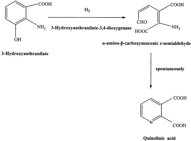
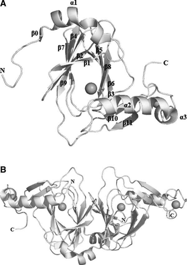
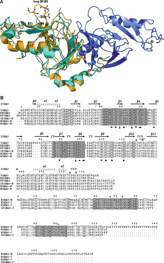
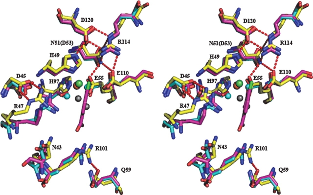
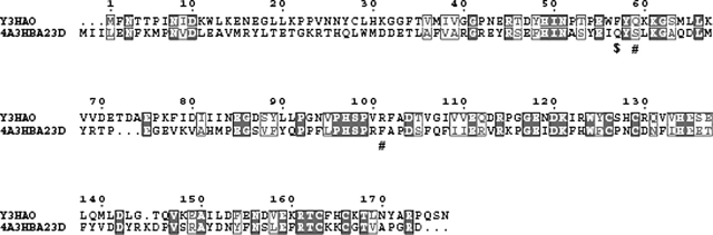
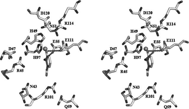
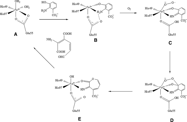
3-Hydroxyanthranilic acid 3,4-dioxygenase (3HAO) is a non-heme ferrous extradiol dioxygenase in the kynurenine pathway from tryptophan. It catalyzes the conversion of 3-hydroxyanthranilate (HAA) to quinolinic acid (QUIN), an endogenous neurotoxin, via the activation of N-methyl-D-aspartate (NMDA) receptors and the precursor of NAD(+) biosynthesis. The crystal structure of 3HAO from S. cerevisiae at 2.4 A resolution shows it to be a member of the functionally diverse cupin superfamily. The structure represents the first eukaryotic 3HAO to be resolved. The enzyme forms homodimers, with two nickel binding sites per molecule. One of the bound nickel atoms occupies the proposed ferrous-coordinated active site, which is located in a conserved double-strand beta-helix domain. Examination of the structure reveals the participation of a series of residues in catalysis different from other extradiol dioxygenases. Together with two iron-binding residues (His49 and Glu55), Asp120, Asn51, Glu111, and Arg114 form a hydrogen-bonding network; this hydrogen-bond network is key to the catalysis of 3HAO. Residues Arg101, Gln59, and the substrate-binding hydrophobic pocket are crucial for substrate specificity. Structure comparison with 3HAO from Ralstonia metallidurans reveals similarities at the active site and suggests the same catalytic mechanism in prokaryotic and eukaryotic 3HAO. Based on sequence comparison, we suggest that bicupin of human 3HAO is the first example of evolution from a monocupin dimer to bicupin monomer in the diverse cupin superfamilies. Based on the model of the substrate HAA at the active site of Y3HAO, we propose a mechanism of catalysis for 3HAO.


Figures

Scheme 1
Scheme 1
The reaction catayzed by 3HAO
Figure 1
Figure 1
According to the gene sequence, residue 47 is reported to be a glycine. The stereo view of the 2Fo-Fc electron density map contoured at 1σ clearly indicates an aspartate in this position. The map shown in green was superimposed on the refined 2.4 Å resolution coordinates of Y3HAO. Carbon (yellow), oxygen (red), and nitrogen (cyan) atoms are shown.
Figure 2
Figure 2
Molecular structure. (A) Ribbon diagram of the Y3HAO subunit. The metal atoms are shown as black spheres. (B) A top view down the axis of the β-barrel of the Y3HAO dimer. The N-terminal strand can be seen to interact with the β-sheet of the other dimer-related subunit. Images here and in the following figures are generated using PYMOL (http://www.pymol.org).
Figure 3
Figure 3
Metal binding site (Ni2+ in the crystal but Fe2+ in vivo) of subunit A of Y3HAO and Fe3+ binding site of RM3HAO. The Ni2+ ion is coordinated by the Nδ1 atom of His49, two Oɛ atoms of Glu55, the Nɛ2 atom of His97 (dashed lines), and two water molecules (W, dashed lines). The Fe3+ ion is coordinated by the Nδ1 atom of His51, two Oɛ atoms of Glu57, the Nɛ2 atom of His95, and two water molecules. The distances between atoms in Å are also shown. The coordinating residues are represented in ball-and-stick models.
Figure 4
Figure 4
(A) Superimpositioning of the Cα backbones of subunit A of Y3HAO (yellow) and RM3HAO (cyan) (PDB code 1YFU; Zhang et al. 2005). Subunit B of Y3HAO is shown in violet. The view is related by ∼25° rotation about the horizontal axis of Figure 2B. (Green spheres) The Ni2+ ions in Y3HAO; (red spheres) iron atoms in RM3HAO. The residues coordinating metal ions, three acid residues in loop β5–β6 of Y3HAO, and one acid residue in the same loop of RM3HAO are shown as ball-and-stick. (Red dashed lines) Hydrogen bonds between Ser88 and His30 and Trp56 of the other dimer-related subunit in Y3HAO. (B) Structure-based sequence alignment of Y3HAO with its homologs. Y3HAO from Saccharomyces cerevisiae; RM3HAO from Ralstonia metallidurans; PF3HAO from Pseudomonas fluorescens; SD3HAO-N, N-terminal domain from Suberites domuncula; SD3HAO-C, C-terminal domain from Suberites domuncula; M3HAO-N, N-terminal domain from Mus musculus; M3HAO-C, C-terminal domain from Mus musculus; H3HAO-N, N-terminal domain from Homo sapiens; H3HAOC, C-terminal domain from Homo sapiens. The secondary structure and residue numbering for Y3HAO are shown above its sequence. (Arrows) β-strands; (large coils) α-helices; (η1) 310 helices. Fully conserved residues (white letters on black background) and conservatively substituted residues (black letters in black boxes) across this group of 3HAOs are indicated. (Black star) Residue in alternate conformations. The image was generated using the program ESPript (Gouet et al. 1999), with secondary structure elements assigned based on 1ZVF for Y3HAO. (Black triangle) Residues binding active-site iron ion; (black rectangles) residues forming the hydrogen-bonding network; (black circle) residues binding two oxygens; (black blob) residues involved in binding the substrate; (black diamond) residues forming hydrophobic pocket; (number sign) conserved cysteines in the monocupin 3HAOs; β0, β1, β2, β4, β7, β9 involved in dimerization are shown. The two characteristic conserved cupin sequences are shaded. Proposed metal coordinating residues of N-terminal domain of H3HAO are indicated with *.
Figure 5
Figure 5
Stereo view of superposition of the active sites of subunit A of Y3HAO (yellow), RM3HAO (PDB code 1YFU, cyan), and the complex structures of the RM3HAO with the inhibitor ClHAA (magenta) and molecular oxygen (red) (PDB code 1YFW, magenta). The metal ions are shown in Y3HAO (green) and RM3HAO (red). The water molecules are shown in Y3HAO (gray) and RM3HAO (cyan). The residue numbering is that of Y3HAO except that E110 is the residue in RM3HAO. Conservative substitution residue in RM3HAO is given in parentheses. D120, N51, E111, and R114, together with H49 and E55, form a hydrogen-bonding network. D47 keeps two hydrogen bonds with Arg45, and Q59 forms a hydrogen bond with R101. Hydrogen bonds are shown as red broken lines.
Figure 6
Figure 6
Sequence alignment between Y3HAO and 4A3HBA23D from Bordetella sp. 10d. The image was generated using the program ESPript (Gouet et al. 1999). Fully conserved residues (white letters on black background) and conservatively substituted residues (black letters in black boxes) are shown. (#) Residues Arg101 and Gln59 are replaced by Phe101 and Ser62 in Bordetella sp. 10d; ($) Phe57, hydrophobic residue lining the substrate, replaced by hydrophilic residue Gln60 in Bordetella sp. 10d.
Figure 7
Figure 7
Model of the substrate HAA at the active site of subunit A of Y3HAO based on the crystal structure of the substrate-bound RM3HAO (PDB code 1YFY) as reported by Zhang et al. (1995). The Oɛ2 atom of E111 forms a hydrogen bond with the C-3 hydroxyl group of HAA, and the Oɛ1 atom of Glu55 forms a hydrogen bond with the C-2 amino group of HAA. These two hydrogen bonds are shown as black broken lines.
Figure 8
Figure 8
The proposed reaction mechanism catalyzed by 3HAO.

Similar articles

Cited by

References

    1. Anand R., Dorrestein P.C., Kinsland C., Begley T.P., Ealick S.E. 2002. Structure of oxalate decarboxylase from Bacillus subtilis at 1.75 Å resolution Biochemistry 41: 7659–7669. - PubMed
    1. Bugg T.D.H. and Lin G. 2001. Solving the riddle of the intradiol and extradiol catechol dioxygenases: How do enzymes control hydroperoxide rearrangements? Chem. Commun. 2001: 941–952.
    1. Bugg T.D., Sanvoisin J., Spence E.L. 1997. Exploring the catalytic mechanism of the extradiol catechol dioxygenases Biochem. Soc. Trans. 25: 81–85. - PubMed
    1. Calderone V., Trabucco M., Menin V., Negro A. 2002. Cloning of human 3-hydroxyanthranilic acid dioxygenase in Escherichia coli: Characterisation of the purified enzyme and its in vitro inhibition by Zn2+ Biochim. Biophys. Acta 1596: 283–292. - PubMed
    1. Cleasby A., Wonacott A., Skarzynski T., Hubbard R.E., Davies G.J., Proudfoot A.E., Bernard A.R., Payton M.A., Wells T.N. 1996. The x-ray crystal structure of phosphomannose isomerase from Candida albicans at 1.7 angstrom resolution Nat. Struct. Biol. 3: 470–479. - PubMed

Publication types

MeSH terms

Substances