. 2006 Apr 28;358(2):559-70.
doi: 10.1016/j.jmb.2006.02.028. Epub 2006 Feb 28.

Structure and substrate recognition of the Escherichia coli DNA adenine methyltransferase

Affiliations

Structure and substrate recognition of the Escherichia coli DNA adenine methyltransferase

John R Horton et al. J Mol Biol. .

Abstract

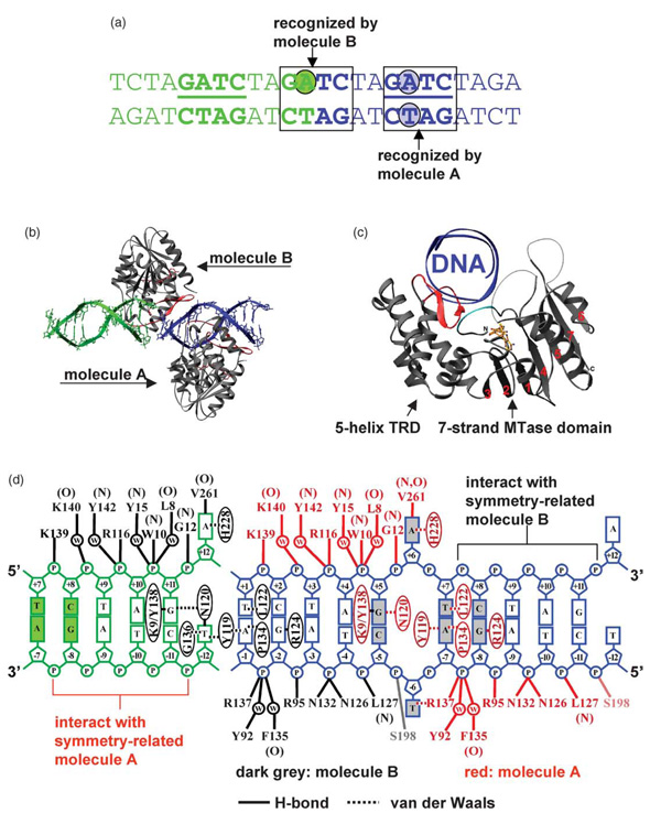
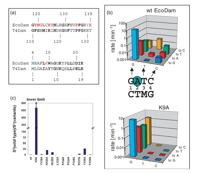
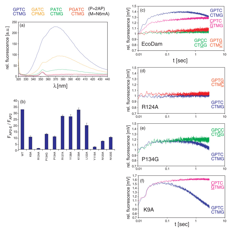
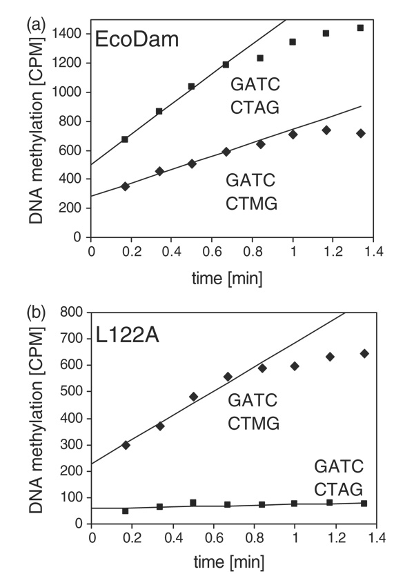
The structure of the Escherichia coli Dam DNA-(adenine-N6)-methyltransferase in complex with cognate DNA was determined at 1.89 A resolution in the presence of S-adenosyl-L-homocysteine. DNA recognition and the dynamics of base-flipping were studied by site-directed mutagenesis, DNA methylation kinetics and fluorescence stopped-flow experiments. Our data illustrate the mechanism of coupling of DNA recognition and base-flipping. Contacts to the non-target strand in the second (3') half of the GATC site are established by R124 to the fourth base-pair, and by L122 and P134 to the third base-pair. The aromatic ring of Y119 intercalates into the DNA between the second and third base-pairs, which is essential for base-flipping to occur. Compared to previous published structures of bacteriophage T4 Dam, three major new observations are made in E.coli Dam. (1) The first Gua is recognized by K9, removal of which abrogates the first base-pair recognition. (2) The flipped target Ade binds to the surface of EcoDam in the absence of S-adenosyl-L-methionine, which illustrates a possible intermediate in the base-flipping pathway. (3) The orphaned Thy residue displays structural flexibility by adopting an extrahelical or intrahelical position where it is in contact to N120.


Figures

Figure 1
Figure 1. Structure of the EcoDam–AdoHcy–12mer DNA complex
(a) Two DNA duplexes (green and blue) are stacked head-to-end, with one GATC site in the middle of each duplex and one in the joint of two duplexes. The nucleotides in extrahelical positions are in shaded circles. (b) Molecule A binds to the GATC site in the middle of each DNA duplex, while EcoDam molecule B binds to the joint of two DNA duplexes. (c) EcoDam contains two domains: a seven-stranded catalytic domain that harbors the binding site for AdoHcy (in stick model) and a DNA-binding domain consisting of a five-helix bundle and a β-hairpin (red) that is conserved in the family of GATC-related MTase orthologs. The N-terminal residues 7 to 10, colored in cyan, also interact with the DNA (see (d)). (D) Summary of the protein–DNA contacts of molecule A (red) and molecule B (grey). Backbone-mediated interactions are indicated with main chain amide (N) or carbonyl (O). For simplicity, only single water molecule (w)-mediated interactions are shown. Focusing on a single DNA duplex (blue), 20 out of 22 phosphate groups interact with three EcoDam molecules (A, B, and symmetry-related molecule B). Thus, the choice of the length (12 base-pairs) and the end sequence of the oligonucleotide used for crystallization optimally maximized the DNA–protein interactions and DNA-mediated protein–protein interactions in the crystal lattice of packing. The only two phosphate groups that are not involved in EcoDam interactions are the 5′ phosphate groups of the two Thy of the central GATC site, which are the phosphate groups missing from the joint GATC site.
Figure 2
Figure 2. EcoDam–DNA base interactions
(a) The target Ade is bound in an alternative nucleotide-binding site, on the outside edge of the active-site pocket formed by the DPPY motif (left panel). The target Ade is superimposed with an omit (base and ribose) electron density map contoured at 3.5σ above the mean (middle panel). Large rotations about the P–O5′ bond of the DNA backbone drive the insertion of Ade into the active site (right panel). The transferable methyl group, modeled onto the sulfur atom of AdoHcy, would lie out of the plane of the Ade base, consistent with the target nitrogen lone pair deconjugated and positioned for an in-line direct methyl group transfer (indicated by an arrow), as seen in the M.TaqI–DNA complex. (b) The hairpin loop of molecule A (red) in the major groove of the blue DNA duplex with a central GATC site. (c) Interaction with the first base-pair (G:C) of GATC. Dotted lines indicate hydrogen bonds. (d) The flipped orphan Thy, superimposed with an omit electron density map contoured at 3.5σ above the mean, stacked with the side-chain of R137. (e) Interaction with the third base-pair (T:A) of GATC. A methyl group is modeled onto the exocyclic amino nitrogen N6 atom of the Ade in the non-target strand. Double arrows indicate van der Waals contacts. (f) Interaction with the fourth base-pair (C:G) of GATC. (G) The orphan Thy–N120 interaction in the joint of two DNA duplexes. The Thy–N120 interaction is similar to other protein side-chain-orphaned base interactions of base-flipping enzymes, such as those for Thy–S112 of T4Dam and Gua-Q237 of M.HhaI. (h) The hairpin loop of molecule B (red) in the joint of two DNA molecules (green and blue). The interactions with the first, third, and fourth bases-pairs are identical with that of molecule A (see (b)).
Figure 3
Figure 3. Recognition of the first base-pair by N-terminal K9
(a) Pair-wise sequence alignment of EcoDam and T4Dam in two regions: the β hairpin loop and the N-terminal loop. The residues colored in red were targets for site-directed mutagenesis. (b) Specificity profile of EcoDam wild-type (top panel) and the K9A variant (bottom panel). For easy comparison, we duplicate the wild-type panel. The single-turn-over methylation rates of the wild-type and the K9A variant are given for the cognate, hemimethylated GATC substrate (light blue bars) as well as for all nine near-cognate hemimethylated substrates. On the horizontal axis, the three positions of the GATC site that are mutated are given (G=GATC, T=GATC, C=GATC, M=N6 mA). The new base introduced at each position is specified on the right-hand axis. The methylation rates of the respective pair of enzyme and substrate are given on the vertical axis (note the logarithmic scale). (c) Specificity factor (defined in Materials and Methods) of EcoDam variants for recognition of the first position of the GATC sequence (S1). The values are given as relative changes with respect to the wild-type. Because no activity could be detected at near-cognate sites modified at the third or fourth base-pair of GATC with the K9A variant, the S1 factor given here is a lower limit, indicated by the arrow. The specificity factors of wild-type EcoDam and the K9A were calculated using the data given in (b) and (c); the data for all other variants were taken from Horton et al.
Figure 4
Figure 4. Base flipping by EcoDam and its variants
(a) Fluorescence intensities of several DNA substrates in the presence of EcoDam. The Figure displays the fluorescence of 2AP (=P in the labels) at the position of target Ade (blue curve), the orphan Thy (orange curve), the Gua1 position of the first pair (green curve), and the immediate 5′ position to the GATC (red curve). The pink curve displays free DNA (the hemimethylated G-2AP-TC) as a control and the black curve is for free enzyme. (b) Changes of relative fluorescence of hemimethylated G-2AP-TC during binding of EcoDam and its variants. (c) Stopped-flow studies of base-flipping using substrates containing the 2AP at the target position (blue curve) and with three near-cognate substrates that carry a single base-pair substitution at the first (pink curve), third (green curve) or fourth base-pair (red curve) of the recognition site. (d)–(f) Stopped-flow studies of base-flipping with EcoDam variants with various substrates: (d) R124A, (e) P134G, and (f) K9A.
Figure 5
Figure 5. Discrimination between unmethylated and hemimethylated DNA
Methylation of unmethylated (squares) and hemimethylated (diamonds) oligonucleotide substrates by the (a) EcoDam (WT) and (b) L122A variant. These experiments were performed in 50 mM Hepes (pH 7.5), 50 mM NaCl, 1 mM EDTA, 0.5 mM DTT and 0.2 µg/µl of BSA using 0.5 µM DNA, 0.25 µM enzyme and 0.76 µM labeled AdoMet.

References

    1. Jones PA, Takai D. The role of DNA methylation in mammalian epigenetics. Science. 2001;293:1068–1070. - PubMed
    1. Jeltsch A. Beyond Watson and Crick: DNA methylation and molecular enzymology of DNA methyltransferases. ChemBioChem. 2002;3:274–293. - PubMed
    1. Klimasauskas S, Kumar S, Roberts RJ, Cheng X. HhaI methyltransferase flips its target base out of the DNA helix. Cell. 1994;76:357–369. - PubMed
    1. Cheng X, Roberts RJ. AdoMet-dependent methylation, DNA methyltransferases and base flipping. Nucl. Acids Res. 2001;29:3784–3795. - PMC - PubMed
    1. Lacks S, Greenberg B. Complementary specificity of restriction endonucleases of Diplococcus pneumoniae with respect to DNA methylation. J. Mol. Biol. 1977;114:153–168. - PubMed

Publication types

MeSH terms

Substances