. 2006 Mar;5(3):488-98.
doi: 10.1128/EC.5.3.488-498.2006.

Lipid rafts in Cryptococcus neoformans concentrate the virulence determinants phospholipase B1 and Cu/Zn superoxide dismutase

Affiliations

Lipid rafts in Cryptococcus neoformans concentrate the virulence determinants phospholipase B1 and Cu/Zn superoxide dismutase

A Rosemary Siafakas et al. Eukaryot Cell. 2006 Mar.

Abstract

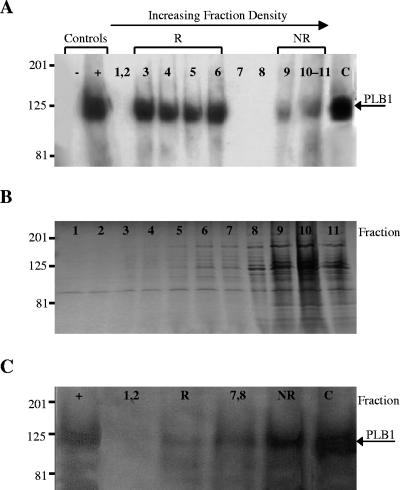
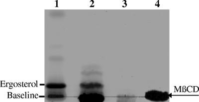
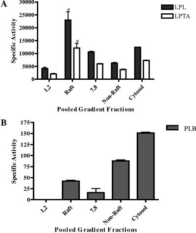
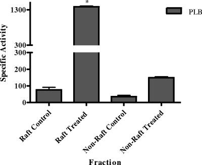
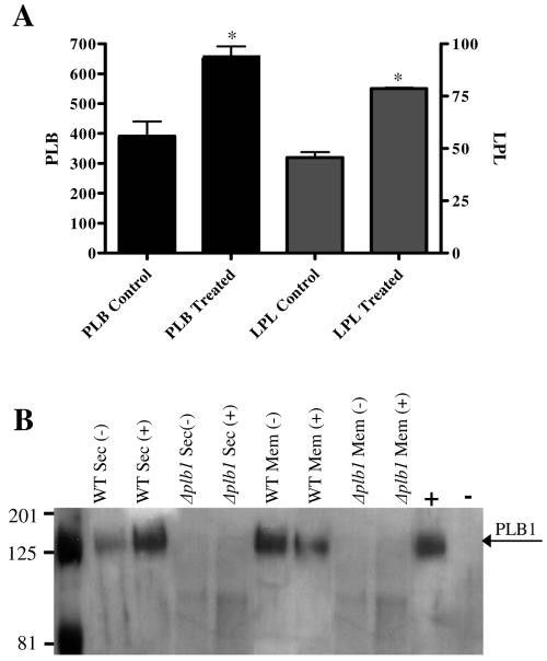
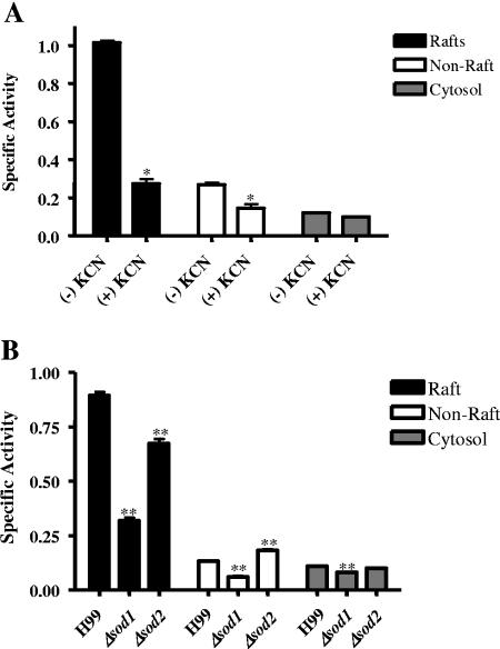
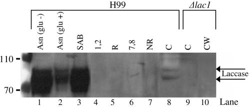
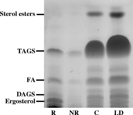
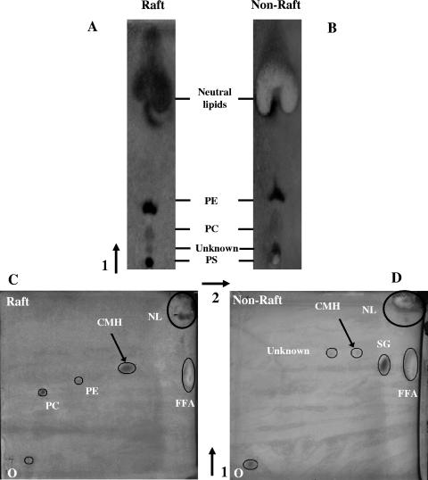
Lipid rafts have been identified in the membranes of mammalian cells, the yeast Saccharomyces cerevisiae, and the pathogenic fungus Candida albicans. Formed by a lateral association of sphingolipids and sterols, rafts concentrate proteins carrying a glycosylphosphatidylinositol (GPI) anchor. We report the isolation of membranes with the characteristics of rafts from the fungal pathogen Cryptococcus neoformans. These characteristics include insolubility in Triton X-100 (TX100) at 4 degrees C, more-buoyant density within a sucrose gradient than the remaining membranes, and threefold enrichment with sterols. The virulence determinant phospholipase B1 (PLB1), a GPI-anchored protein, was highly concentrated in raft membranes and could be displaced from them by treatment with the sterol-sequestering agent methyl-beta-cyclodextrin (MbetaCD). Phospholipase B enzyme activity was inhibited in the raft environment and increased 15-fold following disruption of rafts with TX100 at 37 degrees C. Treatment of viable cryptococcal cells in suspension with MbetaCD also released PLB1 protein and enzyme activity, consistent with localization of PLB1 in plasma membrane rafts prior to secretion. The antioxidant virulence factor Cu/Zn superoxide dismutase (SOD1) was concentrated six- to ninefold in raft membrane fractions compared with nonraft membranes, whereas the cell wall-associated virulence factor laccase was not detected in membranes. We hypothesize that raft membranes function to cluster certain virulence factors at the cell surface to allow efficient access to enzyme substrate and/or to provide rapid release to the external environment.


Figures

FIG. 1.
FIG. 1.
Distribution of PLB1 and total protein in fractionated cryptococcal membranes. Total membranes from C. neoformans were prepared and fractionated by sucrose density gradient centrifugation as described in Materials and Methods. Proteins from the 11 membrane fractions and cytosol were subjected to SDS-PAGE and either transferred to a polyvinylidene difluoride membrane and probed with anti-PLB1 antibody (A) or stained with Coomassie brilliant blue for total protein (B). PLB1 protein distributions in Western blots of membranes prepared from cryptococcal cells treated with MβCD are shown in panel C. Secretions from the wild type (H99) and from Δplb1 were used as positive (+) and negative (−) controls, respectively. Fractionated membranes were pooled as follows, starting from the top of the gradient: fractions 1 and 2, raft fractions 3 to 6 (R), intermediate fractions 7 and 8, nonraft fractions 9 to 11 (NR), and cytosol (C). Gradient fractions are shown from least (left) to most (right) dense, as indicated by the arrow at the top (this applies to panels A, B, and C). Molecular mass standards are indicated (in kilodaltons), and the arrows on the right indicate the position of PLB1.
FIG. 2.
FIG. 2.
Extraction of sterols from cryptococcal cells with MβCD. Cells were incubated with or without 10 mM MβCD for 1 h and pelleted by centrifugation. Supernatants were collected and subjected to lipid extraction as described in Materials and Methods. Extracted lipids were fractionated by TLC and stained for neutral lipids. A lipid band comigrated with the ergosterol standard (lane 1) when MβCD was present in the cell incubation buffer (lane 2) but not when it was absent (lane 3). MβCD that had been taken through the extraction procedure in the absence of cells appears as an intense band on the baseline in lane 4 and is also present in lane 2.
FIG. 3.
FIG. 3.
Distributions of phospholipase activity in fractionated cryptococcal membranes. Total membranes from C. neoformans were prepared, fractionated by sucrose density gradient centrifugation, pooled as described in the legend to Fig. 1, and assayed for LPL/LPTA (A) and PLB (B) activities. Activities are expressed as micromoles of LysoPC substrate degraded (LPL) or DPPC formed (LPTA) per minute per milligram of protein (A) or nanomoles of DPPC degraded (PLB) per minute per milligram of protein (B). *, the increase in LPL/LPTA activity of rafts is statistically significant relative to that of nonraft membranes. P < 0.05 (using an unpaired, nonparametric t test).
FIG. 4.
FIG. 4.
Comparison of PLB activities in intact and disrupted raft membranes. Pooled gradient fractions containing raft and nonraft membranes were incubated with TX100 at 37°C for 1 h to disrupt membrane lipids and then assayed for PLB activity. Activity is expressed as nanomoles of DPPC degraded per minute per milligram of protein. The increase in the activity derived from the treated raft membranes was statistically significant relative to both the untreated raft membrane control and the treated nonraft membranes. P < 0.05 (using an unpaired, nonparametric t test).
FIG. 5.
FIG. 5.
Effects of sterol depletion on PLB1 secretion and membrane association. Cells, either untreated (control) or treated with MβCD for 1 h at 37°C, were washed and resuspended in secretion buffer. At 4 h, cells were pelleted by low-speed centrifugation and the secretions were assayed for PLB and LPL/LPTA specific activities (A). Specific activity is expressed as nanomoles of DPPC formed (PLB) or micromoles of LysoPC (LPL) degraded per minute per milligram of protein. Data are means and standard errors of triplicate assays (three experiments). Asterisks indicate that the increase in specific activity, relative to the respective control, was statistically significant (P < 0.05) using a two-tailed unpaired parametric t test. LPTA activity is not shown, but the percent increase in MβCD-released activity was similar to that obtained for LPL and PLB. Western analysis of secreted (Sec) and membrane (Mem)-associated PLB1 before (−) and after (+) MβCD treatment is shown in panel B. No bands were observed in either fraction from Δplb1. The arrow indicates the position of the PLB1 band. Secretions from the wild-type (H99) and Δplb1 strains were used as positive (+) and negative (−) controls, respectively.
FIG. 6.
FIG. 6.
Membrane distribution of cryptococcal SOD activity. Pooled raft and nonraft membrane fractions and cytosol were prepared from the wild type (H99) and assayed in the presence (+) and absence (−) of KCN, an inhibitor of Cu/Zn SOD (A). Subcellular fractions from H99, Δsod1, and Δsod2 strains were assayed for SOD (B). Data are means and standard errors of the means of triplicate assays (three experiments). *, significant decrease in activity of the wild type in the presence of KCN compared with untreated cells; **, significant change in activity of deletion mutants compared with wild-type cells. One unit of SOD was defined as a 50% decrease in activity relative to controls.
FIG. 7.
FIG. 7.
Subcellular distribution of laccase.β-Glucanase-treated cell wall preparations (lanes 1 to 3 and 10), pooled gradient fractions (lanes 4 to 7), and cytosol (C) (lanes 8 and 9), prepared from either H99 or Δlac1 as indicated in the figure, were subjected to SDS-PAGE and Western blotting with a monoclonal antibody against laccase. Control conditions on the Western blot showed that laccase expression is repressed when glucose is present (lane 2). Arrows indicate the positions of bands attributable to products of the lac1 gene.
FIG. 8.
FIG. 8.
Neutral lipid composition of subcellular fractions from C. neoformans. Lanes were loaded with equal volumes of total lipids from raft fractions (R), nonraft fractions (NR), cytosol (C), and lipid droplets (LD). Lipid separation was achieved with a petroleum ether-diethyl ether-acetic acid (80:20:1 [vol/vol/vol]) solvent system. Separated lipids were visualized with iodine vapor and then sterol spray, followed by charring to reveal all lipid species. Lipids were identified by comparing Rf values with authentic standards. These plates are representative of three separations. The positions of TAGs, diacylglycerols (DAGS), fatty acids (FA), sterol esters, and ergosterol are indicated.
FIG. 9.
FIG. 9.
One- and two-dimensional TLC separations of polar lipids from raft and nonraft fractions. The solvent system used for one-dimensional lipid separation in panels A and B was chloroform-methanol-7 M ammonium hydroxide (65:30:4 [vol/vol/vol]). A two-dimensional separation using the above solvent system in the first dimension and chloroform-methanol-acetic acid-water (170:25:25:4 [vol/vol/vol/vol]) in the second dimension is shown for raft (C) and nonraft (D) membranes. Spots were revealed with iodine vapor or Coomassie blue, which stains FFA white. Aminophospholipids (PE and PS) were identified by using ninhydrin, sterols were identified with sterol spray, and glycolipids were identified with orcinol spray (see Materials and Methods). Spots were also identified by comparison with the Rf values of standard lipids run at the same time as the test plates and using the same solvent system. Where no standards were available (SG and CMH), comparison was made with Rf values of total cryptococcal lipids as used in reference . O, origin; PC, phosphatidylcholine; PE, phosphatidylethanolamine; PS, phosphatidylserine; CMH, ceramide monohexosides; SG, steryl glycosides; FFA, free fatty acids; NL, neutral lipids. These plates are representative of two separations. The arrows indicate the direction of solvent flow.

Similar articles

Cited by

References

    1. Bagnat, M., A. Chang, and K. Simons. 2001. Plasma membrane proton ATPase Pma1p requires raft association for surface delivery in yeast. Mol. Biol. Cell 12:4129-4138. - PMC - PubMed
    1. Bagnat, M., S. Keranen, A. Shevchenko, and K. Simons. 2000. Lipid rafts function in biosynthetic delivery of proteins to the cell surface in yeast. Proc. Natl. Acad. Sci. USA 97:3254-3259. - PMC - PubMed
    1. Bourbonnais, R., and M. G. Paice. 1990. Oxidation of non-phenolic substrates. An expanded role for laccase in lignin biodegradation. FEBS Lett. 267:99-102. - PubMed
    1. Brasitus, T. A., and D. Schachter. 1980. Lipid dynamics and lipid-protein interactions in rat enterocyte basolateral and microvillus membranes. Biochemistry 19:2763-2769. - PubMed
    1. Brouwer, M., T. H. Brouwer, W. Grater, J. J. Enghild, and I. B. Thogersen. 1997. The paradigm that all oxygen-respiring eukaryotes have cytosolic CuZn-superoxide dismutase and that Mn-superoxide dismutase is localized to the mitochondria does not apply to a large group of marine arthropods. Biochemistry 36:13381-13388. - PubMed

Publication types

MeSH terms

LinkOut - more resources