. 2006 Mar;5(3):587-600.
doi: 10.1128/EC.5.3.587-600.2006.

Mutational analysis of the glycosylphosphatidylinositol (GPI) anchor pathway demonstrates that GPI-anchored proteins are required for cell wall biogenesis and normal hyphal growth in Neurospora crassa

Affiliations

Mutational analysis of the glycosylphosphatidylinositol (GPI) anchor pathway demonstrates that GPI-anchored proteins are required for cell wall biogenesis and normal hyphal growth in Neurospora crassa

Shaun M Bowman et al. Eukaryot Cell. 2006 Mar.

Abstract

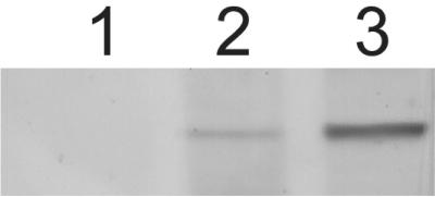
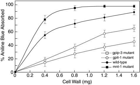
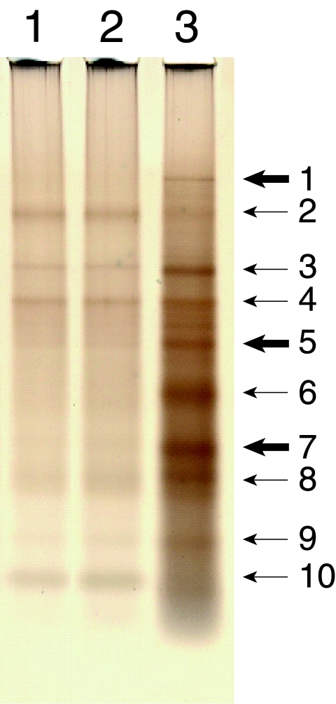
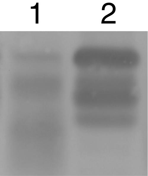
Using mutational and proteomic approaches, we have demonstrated the importance of the glycosylphosphatidylinositol (GPI) anchor pathway for cell wall synthesis and integrity and for the overall morphology of the filamentous fungus Neurospora crassa. Mutants affected in the gpig-1, gpip-1, gpip-2, gpip-3, and gpit-1 genes, which encode components of the N. crassa GPI anchor biosynthetic pathway, have been characterized. GPI anchor mutants exhibit colonial morphologies, significantly reduced rates of growth, altered hyphal growth patterns, considerable cellular lysis, and an abnormal "cell-within-a-cell" phenotype. The mutants are deficient in the production of GPI-anchored proteins, verifying the requirement of each altered gene for the process of GPI-anchoring. The mutant cell walls are abnormally weak, contain reduced amounts of protein, and have an altered carbohydrate composition. The mutant cell walls lack a number of GPI-anchored proteins, putatively involved in cell wall biogenesis and remodeling. From these studies, we conclude that the GPI anchor pathway is critical for proper cell wall structure and function in N. crassa.


Figures

FIG. 1.
FIG. 1.
GPI anchor mutants have altered gross and hyphal morphologies. Panels A to F are photographs of strains that were inoculated on agar growth medium in standard petri dishes. Panels G to L are pictures of the same strains that were inoculated between two cellophane sheets on agar growth medium, and the growing edge of each colony was photographed at a magnification of ×400. All cultures were incubated at room temperature, with the exception of the 34-15 (gpig-1) temperature-sensitive mutant, which was grown at 39°C to induce the mutant phenotype. Colonies of the wild-type (GTH-16) strain (A and G), MSA-7 (gpip-1) mutant (B and H), 34-15 (gpig-1) mutant (C and I), gpip-2 mutant (D and J), gpip-3 mutant (E and K), and gpit-1 mutant (F and L) are shown. The images in panels A to D, H, and J to L were captured at 48 h after inoculation. Panels E and F are shown at 10 days after inoculation. Panel G is shown at 24 h after inoculation. For the micrograph in panel I, the 34-15 (gpig-1) mutant was initially grown at room temperature for 24 h and then shifted to 39°C for an additional 6 h prior to examination. The scale bar in panel L represents a distance of 10 μm.
FIG. 2.
FIG. 2.
The GPIP-3 protein is absent in the gpip-3 mutant. Samples of gpip-3 null mutant (lane 1) and wild-type (GTH-16) (lane 2) total cellular extracts and a wild-type (GTH-16) membrane preparation (lane 3) were separated by SDS-PAGE and analyzed by Western blotting using an anti-GPIP-3 antibody.
FIG. 3.
FIG. 3.
The gpip-3 and gpit-1 mutants have an abnormal “cell-within-a-cell” morphology. The gpip-3 mutant, gpit-1 mutant, and wild-type (GTH-16) strains were grown atop cellophane sheets on standard agar growth medium and prepared for electron microscopy as described in Materials and Methods. Representative electron micrographs of a wild-type cell (A), the gpip-3 mutant (B), and the gpit-1 mutant (C) are shown. Note the “cell-within-a-cell” morphology characteristic of the mutant cells. The scale bar in panel C represents a distance of 1 μm.
FIG. 4.
FIG. 4.
Cell wall absorption of aniline blue dye. Purified cell walls were prepared from the gpip-3 and gpit-1 mutants, the mnt-1 mutant, and the wild-type (GTH-16) parental strain as described in Materials and Methods. Increasing amounts of the mutant and wild-type cell wall preparations were incubated with a solution of 0.002% aniline blue dye, and the amount of aniline blue dye absorbed by each was determined as described in Materials and Methods. Graphed values are the means ± standard deviations of the results from three independent determinations.
FIG. 5.
FIG. 5.
The gpig-1 mutant lacks a number of “cell wall-associated” proteins at the restrictive temperature. The gpig-1 temperature-sensitive mutant and wild-type strain were grown at the permissive (22°C) and restrictive (39°C) temperatures and harvested, and cell extracts of each were prepared. Aliquots of the SDS-soluble material from each cell extract were separated by SDS-PAGE and analyzed by Western blotting using an “anti-cell wall” antibody. Samples of the gpig-1 mutant at 22°C (lane 1) and 39°C (lane 2) and the wild-type strain at 22°C (lane 3) and 39°C (lane 4) are shown. The molecular masses indicated at the right are in kilodaltons.
FIG. 6.
FIG. 6.
The gpip-3 and gpit-1 mutants have altered “integral cell wall” protein compositions. Purified cell walls from the gpip-3 and gpit-1 mutants and the wild-type (GTH-16) strain were digested with TFMS acid as described in Materials and Methods. The total protein released from 1 mg of starting cell wall material from the gpip-3 mutant (lane 1), gpit-1 mutant (lane 2), and wild-type strain (lane 3) were separated by SDS-PAGE and visualized by silver staining. The 10 major protein bands from the wild-type cell wall are indicated with arrows. Those bands containing GPI-anchored proteins are highlighted with thick arrows. See Table 1 for a listing of both the GPI-anchored and nonanchored proteins detected in the major protein bands.
FIG. 7.
FIG. 7.
The gpip-3 mutant has reduced levels of ACW-1. Samples of gpip-3 null mutant (lane 1) and wild-type (GTH-16) (lane 2) total cellular extracts were separated by SDS-PAGE and analyzed by Western blotting using an anti-ACW-1 antibody.

References

    1. Akins, R. A., and A. M. Lambowitz. 1985. General method for cloning Neurospora crassa nuclear genes by complementation of mutants. Mol. Cell. Biol. 5:2272-2278. - PMC - PubMed
    1. Alloush, H. M., J. L. Lopez-Ribot, B. J. Masten, and W. L. Chaffin. 1997. 3-Phosphoglycerate kinase: a glycolytic enzyme present in the cell wall of Candida albicans. Microbiology 143:321-330. - PubMed
    1. Angiolella, L., M. Facchin, A. Stringaro, B. Maras, N. Simonetti, and A. Cassone. 1996. Identification of a glucan-associated enolase as a main cell wall protein of Candida albicans and an indirect target of lipopeptide antimycotics. J. Infect. Dis. 173:684-690. - PubMed
    1. Benachour, A., G. Sipos, I. Flury, F. Reggiori, E. Canivenc-Gansel, C. Vionnet, A. Conzelmann, and M. Benghezal. 1999. Deletion of GPI7, a yeast gene required for addition of a side chain to the glycosylphosphatidylinositol (GPI) core structure, affects GPI protein transport, remodeling, and cell wall integrity. J. Biol. Chem. 274:15251-15261. - PubMed
    1. Benghezal, M., P. N. Lipke, and A. Conzelmann. 1995. Identification of six complementation classes involved in the biosynthesis of glycosylphosphatidylinositol anchors in Saccharomyces cerevisiae. J. Cell Biol. 130:1333-1344. - PMC - PubMed

Publication types

MeSH terms

LinkOut - more resources