Review
. 2006 Mar;70(1):177-91.
doi: 10.1128/MMBR.70.1.177-191.2006.

The where, when, and how of organelle acidification by the yeast vacuolar H+-ATPase

Affiliations
Review

The where, when, and how of organelle acidification by the yeast vacuolar H+-ATPase

Patricia M Kane. Microbiol Mol Biol Rev. 2006 Mar.

Abstract

All eukaryotic cells contain multiple acidic organelles, and V-ATPases are central players in organelle acidification. Not only is the structure of V-ATPases highly conserved among eukaryotes, but there are also many regulatory mechanisms that are similar between fungi and higher eukaryotes. These mechanisms allow cells both to regulate the pHs of different compartments and to respond to changing extracellular conditions. The Saccharomyces cerevisiae V-ATPase has emerged as an important model for V-ATPase structure and function in all eukaryotic cells. This review discusses current knowledge of the structure, function, and regulation of the V-ATPase in S. cerevisiae and also examines the relationship between biosynthesis and transport of V-ATPase and compartment-specific regulation of acidification.


Figures
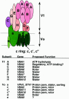
FIG. 1.
FIG. 1.
Subunit composition and structural model of the yeast V-ATPase. V1 subunits are shown in pink and purple and V0 subunits are shown in green. A single stator stalk containing subunits C, E, G, and H is shown, but recent electron microscopy evidence suggests that there may be two peripheral stalks in the closely related Neurospora crassa V-ATPase (152). If there are two stator stalks in the yeast enzyme, it is likely that each stalk will contain subunits E and G, but one stalk may contain subunit H, and the other subunit C (152).
FIG. 2.
FIG. 2.
Assembly and trafficking of the yeast V-ATPase. Possible steps in assembly and transport of Vph1p-containing and Stv1p-containing V-ATPases are shown here and described in more detail in the text. Shading of different organelles indicates the extent of acidification in that compartment; the vacuole is most intensely colored as the most acidic compartment in the yeast cell, and the first compartment in the secretory pathway showing any evidence of acidification in yeast is the Golgi apparatus. Vph1p-containing V-ATPases are known to travel to the vacuole via the prevacuolar compartment (PVC) (117) and are believed to reach the prevacuolar compartment via the early endosome, which is also likely to be somewhat acidic (134). Stv1p-containing V-ATPases appear to cycle between the prevacuolar compartment and the vacuole (68), and may travel through the early endosome as well. The RAVE complex is known to assist in reassembly of V1 and V0 complexes at the vacuole (131), and may also assist in assembly at the early endosome (134).
FIG. 3.
FIG. 3.
Overview of reversible disassembly in yeast cells. Intact, active V-ATPase complexes are rapidly disassembled into free V1 and V0 complexes in response to glucose deprivation (61). Glucose readdition results in reassembly of the V-ATPase, and is slow and incomplete in the absence of the RAVE complex (131). RAVE binding to V1 is compromised in mutants lacking subunit E or G, suggesting that these two subunits are involved in the interaction of V1 and RAVE (135). Subunits that are believed to undergo major conformational changes during disassembly are identified: the H subunit shifts from being an activator of V1V0 complexes to being an inhibitor of free V1 complexes (113), the cytoplasmic N-terminal domain of the a subunit appears to fold down onto the rest of V0 (157), and the C subunit is released from both the V1 and the V0 sectors during disassembly (61).

Similar articles

Cited by

References

    1. Abazeed, M. E., J. M. Blanchette, and R. S. Fuller. 2004. Cell-free transport from the TGN to late endosome requires factors involved in formation and consumption of clathrin-coated vesicles. J. Biol. Chem. 280:4442-4450. - PubMed
    1. Aksimentiev, A., I. A. Balabin, R. H. Fillingame, and K. Schulten. 2004. Insights into the molecular mechanism of rotation in the F0 sector of ATP synthase. Biophys. J. 86:1332-1344. - PMC - PubMed
    1. Arai, H., S. Pink, and M. Forgac. 1989. Interaction of anions and ATP with the coated vesicle proton pump. Biochemistry 28:3075-3082. - PubMed
    1. Arata, Y., J. D. Baleja, and M. Forgac. 2002. Localization of subunits D, E, and G in the yeast V-ATPase complex using cysteine-mediated cross-linking to subunit B. Biochemistry 41:11301-11307. - PubMed
    1. Bauerle, C., M. N. Ho, M. A. Lindorfer, and T. H. Stevens. 1993. The Saccharomyces cerevisiae VMA6 gene encodes the 36-kDa subunit of the vacuolar H+-ATPase membrane sector. J. Biol. Chem. 268:12749-12757. - PubMed

Publication types

MeSH terms

Substances

LinkOut - more resources