Review
. 2006 Mar;70(1):192-221.
doi: 10.1128/MMBR.70.1.192-221.2006.

Sortases and the art of anchoring proteins to the envelopes of gram-positive bacteria

Affiliations
Review

Sortases and the art of anchoring proteins to the envelopes of gram-positive bacteria

Luciano A Marraffini et al. Microbiol Mol Biol Rev. 2006 Mar.

Abstract

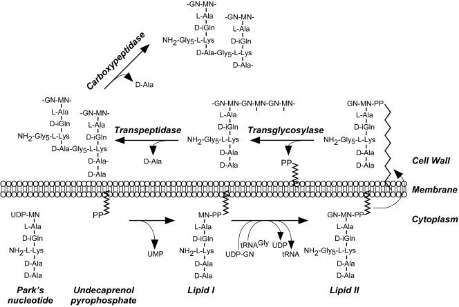
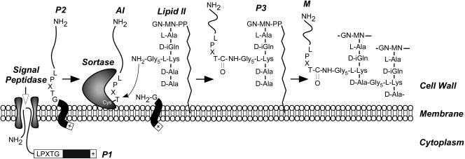
The cell wall envelopes of gram-positive bacteria represent a surface organelle that not only functions as a cytoskeletal element but also promotes interactions between bacteria and their environment. Cell wall peptidoglycan is covalently and noncovalently decorated with teichoic acids, polysaccharides, and proteins. The sum of these molecular decorations provides bacterial envelopes with species- and strain-specific properties that are ultimately responsible for bacterial virulence, interactions with host immune systems, and the development of disease symptoms or successful outcomes of infections. Surface proteins typically carry two topogenic sequences, i.e., N-terminal signal peptides and C-terminal sorting signals. Sortases catalyze a transpeptidation reaction by first cleaving a surface protein substrate at the cell wall sorting signal. The resulting acyl enzyme intermediates between sortases and their substrates are then resolved by the nucleophilic attack of amino groups, typically provided by the cell wall cross bridges of peptidoglycan precursors. The surface protein linked to peptidoglycan is then incorporated into the envelope and displayed on the microbial surface. This review focuses on the mechanisms of surface protein anchoring to the cell wall envelope by sortases and the role that these enzymes play in bacterial physiology and pathogenesis.


Figures

FIG. 1.
FIG. 1.
Peptidoglycan synthesis in S. aureus. Park's nucleotide, a soluble nucleotide precursor, originates in the bacterial cytoplasm by successive addition of l-stereoisomer amino acids (l-Ala and l-Lys) as well as d-stereoisomer amino acids (d-isoglutamine [d-iGln] and d-Ala) to UDP-N-acetylmuramic acid (UDP-NM). Precursor transfer to undecaprenol pyrophosphate, a bacterial membrane carrier, generates lipid I and removes UMP nucleotide. Lipid I modification with N-acetylglucosamine (GN) and pentaglycine cross bridge formation at the ɛ-amino of l-Lys with tRNAGly substrate generates lipid II. Following translocation across the cytoplasmic membrane, lipid II serves as substrate for PBPs that catalyze three reactions: transglycosylation, transpeptidation, and carboxypeptidation. Transglycosylases polymerize MN-GN subunits into repeating disaccharide chains, the glycan strands. Transpeptidases cleave the amide bond of the terminal d-Ala in pentapeptide precursors and generate an amide bond between the carboxyl group of d-Ala at position four and the amino group of pentaglycine cross bridges in wall peptides. Carboxypeptidases hydrolyze the C-terminal d-Ala of most non-cross-linked pentapeptides to yield mature peptidoglycan.
FIG. 2.
FIG. 2.
Sortase A-dependent surface display of staphylococcal proteins. Sortase is responsible for the anchoring of 20 different surface proteins to the cell wall of S. aureus strain Newman. One of these surface proteins, protein A, binds to the Fc terminus of mammalian immunoglobulins in a nonimmune fashion, causing decoration of the staphylococcal surface with antibody. Using Cy3-conjugated immunoglobulin and S. aureus strain Newman, protein A display on the bacterial surface was revealed with phase-contrast microscopy and fluorescence microscopy. Protein A display on the staphylococcal surface is abrogated in the srtA mutant strain (SKM3).
FIG. 3.
FIG. 3.
Cell wall anchor structure of staphylococcal surface proteins. The C-terminal threonine of surface proteins, generated by sortase A-mediated cleavage between the threonine and the glycine of the LPXTG motif, is amide linked to the pentaglycine cross bridge of S. aureus cell wall peptidoglycan. Treatment of the staphylococcal peptidoglycan with lysostaphin (glycyl-glycine endopeptidase), mutanolysin [N-acetylmuramidase that cleaves the β(1-4) O-glycosidic bond between N-acetylmuramic acid and N-acetylglucosamine (GN)], amidase (N-acetylmuramoyl-l-Ala amidase), or Φ11 hydrolase (N-acetylmuramoyl-l-Ala amidase and d-Ala-Gly endopeptidase) releases surface protein with the predicted C-terminal cell wall anchor structures.
FIG. 4.
FIG. 4.
Structure of S. aureus sortase A bound to the LPETG substrate. Sortase folds into an eight-stranded β-barrel structure. The active site resides in a depression formed by β7 and β8 strands. The side chains of His120, Cys184, and Arg197, all of which are absolutely conserved among sortases and are required for activity, as well as the LPETG substrate are drawn with ball-and-stick structures. Cys184 performs a nucleophilic attack on the peptide bond between the threonine and the glycine residues of the substrate, resulting in the formation of an acyl intermediate with the carboxyl group of the C-terminal threonine thioester linked to the sulfur of Cys184. This intermediate is resolved by a second nucleophilic attack on the thioester bond, which results in the release of the reaction products (the structure was generated from atomic coordinates deposited in Protein Data Bank, PDB ID 1T2P) (227).
FIG. 5.
FIG. 5.
Cell wall sorting pathway of surface proteins in gram-positive bacteria. Surface proteins are first synthesized in the bacterial cytoplasm as full-length precursors (P1) containing an N-terminal signal sequence and a C-terminal sorting signal. The signal sequence directs the cellular export of the polypeptide through the Sec system and, upon translocation, is cleaved by signal peptidase. The product of this reaction, the P2 precursor harboring only the C-terminal sorting signal, is retained within the secretory pathway via its C-terminal hydrophobic domain (black box) and positively charged tail (+). Sortase, a membrane-anchored transpeptidase with active-site cysteine, cleaves the peptide bond between the threonine (T) and the glycine (G) of the LPXTG motif, generating an acyl intermediate (AI). Lipid II, the peptidoglycan biosynthesis precursor, and its pentaglycine cross bridge (Gly5) amino group attack the acyl intermediate, linking the C-terminal threonine of the surface protein to lipid II (P3 precursor) and regenerating the active site of sortase. The P3 precursor functions as a substrate for penicillin binding proteins and is incorporated into the cell wall envelope to generate mature anchored surface protein (M), which is also displayed on the bacterial surface. This pathway is universal in many gram-positive bacteria, and the functional elements of cell wall cross bridges, LPXTG motif, sortase, and penicillin binding proteins are conserved.
FIG. 6.
FIG. 6.
Isd-mediated heme-iron uptake in S. aureus. A. The isd locus is comprised of isdA, isdB, and isdC, which encode cell wall-anchored proteins carrying LPKTG, LPQTG, and NPQTN motifs in their respective sorting signals. Located elsewhere in the S. aureus genome, isdH and isdI encode a fourth LPKTG surface protein and a heme oxygenase, respectively. All isd genes are regulated by the ferric uptake repressor (Fur), which represses transcription under iron-replete conditions by binding to fur boxes present in promoter regions (shaded boxes). Arrows indicate the direction of transcription. B. A model for Isd-mediated heme-iron transport across the cell wall of S. aureus. IsdA, IsdB, and IsdH are anchored to the cell wall by sortase A and function as receptors for hemoprotein ligands, including haptoglobin (Hpt), hemoglobin (Hb), or heme. Upon binding to Isd receptors, heme is released from the hemoproteins by an as-yet-undefined mechanism and passaged through the cell wall in an IsdC-dependent manner. Treatment of staphylococcal cells with extracellular proteinase K completely degrades IsdB, only partially digests IsdA, and leaves IsdC intact, suggesting different degrees of surface exposure for each of these cell wall proteins. The heme molecule is then transported through the membrane transport system composed of IsdDEF into the cytoplasm. Upon entry into the cytoplasm, heme is degraded by IsdG and IsdI heme monooxygenases. This leads to the release of free iron for use by the bacterium as a nutrient source. (Adapted from reference with permission from Elsevier.)
FIG. 7.
FIG. 7.
Cell wall anchor structure of staphylococcal IsdC. The C-terminal threonine of IsdC, generated by sortase B-mediated cleavage between the threonine and the asparagine of the NPQTN motif, is amide linked to the pentaglycine cross bridge of S. aureus cell wall peptidoglycan. Treatment of the staphylococcal peptidoglycan with lysostaphin (glycyl-glycine endopeptidase), mutanolysin [N-acetylmuramidase that cleaves the β(1-4) O-glycosidic bond between N-acetylmuramic acid and N-acetylglucosamine (GN)], amidase (N-acetylmuramoyl-l-Ala amidase), or Φ11 hydrolase (N-acetylmuramoyl-l-Ala amidase and d-Ala-Gly endopeptidase) releases surface protein with the predicted C-terminal cell wall anchor structures. In contrast to sortase A substrates, sortase B-anchored IsdC is attached to only six or seven disaccharide subunits and its wall peptides are either non-cross-linked (murein pentapeptides containing an extra C-terminal d-Ala) or linked to only two or three peptidoglycan subunits.
FIG. 8.
FIG. 8.
Crystal structure of S. aureus sortase B. Sortase A and sortase B fold into very similar β-barrel structures; however, sortase B harbors three α helices that are absent in sortase A (here shown in orange) and that may contribute to the unique properties of sortase B substrate specificity and anchoring. Cys223, His130, and Arg233 are equivalent to sortase A Cys184, His120, and Arg197, respectively, and, along with Asn225, presumably constitute the active site of sortase B (the structure was generated from atomic coordinates deposited in Protein Data Bank, PDB ID 1QXA) (228).
FIG. 9.
FIG. 9.
Corynebacterium diphtheriae pili. A. Genetic organization of the spa locus of C. diphtheriae NCTC13129. Predicted promoters as well as the direction of transcription are shown with arrows. B to D. Corynebacterial pili stained with specific antiserum (anti-SpaA [B], anti-SpaB [C], or anti-SpaC [D]) and IgG-conjugated 12-nm gold particles. Samples were viewed by transmission electron microscopy. Bars indicate a distance of 0.2 μm. (Adapted from reference with permission of Blackwell Publishing.)
FIG. 10.
FIG. 10.
Model for sortase-mediated pilus polymerization in C. diphtheriae. Sortase is thought to catalyze the polymerization of pili on the corynebacterial cell surface. Pilin subunits are typical sortase substrates, containing an N-terminal signal peptide (SP) that promotes secretion through the Sec system and a C-terminal cell wall sorting signal. SpaC is thought to be the first subunit to be incorporated into pili; if so, this might account for the detection of SpaC at the tip of the pilin fiber. The sortase-SpaC acyl intermediate may be attacked by the free amino group of a conserved lysine residue (K) present in the pilin motif of SpaA. The SpaA sorting signal would be in turn cleaved by sortase and linked to the lysine of a second SpaA pilin subunit. The remainder of the filament may then assemble by a sequence of similar transpeptidation reactions, and the polymerized pili may then be transferred to cell wall cross bridges for immobilization in the bacterial envelope. (Adapted from reference with permission from Elsevier.)

Similar articles

Cited by

References

    1. Abrahmsen, L., T. Moks, B. Nilsson, U. Hellman, and M. Uhlen. 1985. Analysis of signals for secretion in the staphylococcal protein A gene. EMBO J. 4:3901-3906. - PMC - PubMed
    1. Akabas, M. H., and A. Karlin. 1995. Identification of acetylcholine receptor channel-lining residues in the M1 segment of the alpha-subunit. Biochemistry 34:12496-12500. - PubMed
    1. Albus, A., R. D. Arbeit, and J. C. Lee. 1991. Virulence studies of Staphylococcus aureus mutants altered in type 5 capsule production. Infect. Immun. 59:1008-1014. - PMC - PubMed
    1. Bae, T., and O. Schneewind. 2003. The YSIRK-G/S motif of staphylococcal protein A and its role in efficiency of signal peptide processing. J. Bacteriol. 185:2910-2919. - PMC - PubMed
    1. Banks, D. J., S. F. Porcella, K. D. Barbian, S. B. Beres, L. E. Philips, J. M. Voyich, F. R. DeLeo, J. M. Martin, G. A. Somerville, and J. M. Musser. 2004. Progress toward characterization of the group A Streptococcus metagenome: complete genome sequence of a macrolide-resistant serotype M6 strain. J. Infect. Dis. 190:727-738. - PubMed

Publication types

MeSH terms

LinkOut - more resources