Review
. 2006 Mar;70(1):222-52.
doi: 10.1128/MMBR.70.1.222-252.2006.

Adaptation and acclimation of photosynthetic microorganisms to permanently cold environments

Affiliations
Review

Adaptation and acclimation of photosynthetic microorganisms to permanently cold environments

Rachael M Morgan-Kiss et al. Microbiol Mol Biol Rev. 2006 Mar.

Abstract

Persistently cold environments constitute one of our world's largest ecosystems, and microorganisms dominate the biomass and metabolic activity in these extreme environments. The stress of low temperatures on life is exacerbated in organisms that rely on photoautrophic production of organic carbon and energy sources. Phototrophic organisms must coordinate temperature-independent reactions of light absorption and photochemistry with temperature-dependent processes of electron transport and utilization of energy sources through growth and metabolism. Despite this conundrum, phototrophic microorganisms thrive in all cold ecosystems described and (together with chemoautrophs) provide the base of autotrophic production in low-temperature food webs. Psychrophilic (organisms with a requirement for low growth temperatures) and psychrotolerant (organisms tolerant of low growth temperatures) photoautotrophs rely on low-temperature acclimative and adaptive strategies that have been described for other low-temperature-adapted heterotrophic organisms, such as cold-active proteins and maintenance of membrane fluidity. In addition, photoautrophic organisms possess other strategies to balance the absorption of light and the transduction of light energy to stored chemical energy products (NADPH and ATP) with downstream consumption of photosynthetically derived energy products at low temperatures. Lastly, differential adaptive and acclimative mechanisms exist in phototrophic microorganisms residing in low-temperature environments that are exposed to constant low-light environments versus high-light- and high-UV-exposed phototrophic assemblages.


Figures

FIG. 1.
FIG. 1.
Oxygenic photosynthetic electron transport chain in the thylakoids of green algae and higher plants. Three major membrane-bound protein complexes functioning in series, photosystem II (PSII), cytochrome b6f complex (Cyt b6f), and photosystem I (PSI), are required to transport electrons from water to NADP+. Light energy is absorbed by the light-harvesting complexes, and excitation energy is transferred to the reaction centers, where it is used to drive charge separation of a chlorophyll pair (P680 and P700 for PSII and PSI, respectively). Electrons are transported from PSII to cytochrome b6f across the thylakoid membrane by the mobile transporter plastoquinone (PQ/PQH2), and from cytochrome b6f to PSI in the lumenal space by the small protein plastocyanin (PC). Electrons flow from NADPH to downstream metabolic reactions such as carbon and nutrient assimilation. QA, quinone A; QB, quinone B; FNR, ferredoxin-NADP oxidoreductase; A0, A1 FX, FA, and FB, intermediate electron acceptors of Photosystem I.
FIG. 2.
FIG. 2.
Schematic showing the function of the photosynthetic electron transport chain as a redox sensor. The process of photosynthesis integrates the fast temperature-independent photochemical reactions of light absorption and charge separation with the “slow” processes of electron transport and downstream utilization of electron sinks through growth and metabolism. Any environmental stress that perturbs the poise between energy absorbed and energy utilized is sensed by the photoautotrophic organism at the level of the redox state of intersystem electron transport pool (excitation pressure, redox) and/or the build-up of protons across the thylakoid membranes (proton motive force). Low temperatures (Low T) cause an imbalance between energy absorbed and energy utilized by reducing rates of energy consumption by downstream metabolic processes. The energy imbalance is corrected at the level of either light absorption via modulation in the absorptive cross-section of PSII (e.g., dissipation of excess energy as heat, NPQ) or at the level of energy utilization (e.g., modulations at the level of Calvin cycle enzymes). Ox, oxidized; red, reduced.
FIG. 3.
FIG. 3.
Map of the McMurdo Dry Valleys, Antarctica. Unusual meteorological conditions in the dry valleys produce the only permanently ice-covered lakes on Earth and the liquid water beneath the ice allows one of the few refugia for microbial communities in continental Antarctica. Each lake exhibits a unique biogeochemistry and therefore a unique microbial consortium. A Chlamydomonas sp. has been identified in many of the lakes. C. raudensis UWO 241 was isolated from the east lobe of Lake Bonney, Taylor Valley.
FIG. 4.
FIG. 4.
Pictorial representation of the major food web components and their linkages within Lake Bonney (Taylor Valley, Antarctica). The rotifer is a Philodina sp., the large ciliate is a Euplotes sp., and the phytoplankton is a Chlamydomonas sp. Note the bacteria attached to the surface of the Chlamydomonas cells which appear as threadlike structures on the upper portion of the cell; flagella are evident on the right apex of the cell. Note the absence of higher trophic levels. (Reprinted from reference with permission of the publisher.)
FIG. 5.
FIG. 5.
Physical, chemical, and biological parameters from the west and east lobes of Lake Bonney. SRP, soluble reactive phosphorus; PPR, phytoplankton primary productivity; Chl-a, chlorophyll a; TdR, bacterial productivity measured as [3H]thymidine incorporation into DNA; PSU, practical salinity units. (Reprinted from reference with permission of the publisher.)
FIG. 6.
FIG. 6.
Underwater PAR plots taken from the east lobe of Lake Bonney over the time period of 1999-2003.
FIG. 7.
FIG. 7.
Vertical profiles of the major phytoplankton species in the east lobe of Lake Bonney at selected time intervals over the past decade.
FIG. 8.
FIG. 8.
Electron micrographs of C. raudensis grown under laboratory-controlled conditions (8°C/20 μmol photons m−2 s−1) showing a single cell (A) and a membrane-bound colony (B). Note starch accumulation within the pyrenoids (P). C, chloroplast; N, nucleus; G, Golgi apparatus; M, mitochondrion; F, flagellum. Scale bars, 1.0 μm. (Reprinted and modified from reference with permission of Blackwell Publishing.)
FIG. 9.
FIG. 9.
Dependence of rates of growth in C. raudensis on temperature (A) and irradiance (B). A. Cultures were grown under low irradiance (20 μmol m−2 s−1) and variable growth temperatures. B. Cultures were grown under optimal growth temperature (8°C) and variable growth irradiances.
FIG. 10.
FIG. 10.
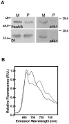
A. Abundance of major thylakoid proteins of photosystems I and II in the mesophilic species C. reinhardtii (M) and the psychrophillic C. raudensis (P) grown at the optimal growth temperature (29 and 8°C, respectively) and low irradiance levels. PsaA/B, photosystem I core reaction center proteins; D1, major photosystem II reaction protein; p18.1 and p22.1, representative light-harvesting I polypeptides. B. Low-temperature 77 K fluorescence emission spectra measured in vivo in C. reinhardtii (dotted line) and C. raudensis (solid line) cultures grown under same conditions as in A. (Modified from reference with kind permission of Springer Science and Business Media.)
FIG. 11.
FIG. 11.
Sequence alignment of cytochrome f proteins from C. raudensis and C. reinhardtii. The protein sequence was predicted from the DNA sequence and aligned using the DNAman program. Black boxes indicate identical amino acids. Green shading, similar amino acids. Black arrow, N terminus of mature cytochrome f protein; red box, heme binding motif; brown box, transmembrane helix; gray underline, C terminus localized in stromal side of thylakoid membrane; blue arrows, lysine residues involved in plastocyanin docking; yellow arrows, amino acid residues involved in water chain formation.
FIG. 12.
FIG. 12.
State transitions in the mesophilic C reinhardtii (left panel) versus the psychrophilic C. raudensis (right panel). A. The 77 K fluorescence emission spectra of whole cells exposed to state I (solid line) or state II (dotted line) conditions. B. Immunoblot of isolated thylakoid membranes with antiphosphothreonine antibodies to detect phosphorylation patterns in proteins of thylakoids exposed to state I (I) or state II (II) conditions. A state I response was induced by exposure of freshly harvested cells from an exponentially growing culture to far-red light for 15 min. A state II response was induced by incubation of samples in the dark under anaerobic conditions. (Modified from reference with kind permission of Springer Science and Business Media.)
FIG. 13.
FIG. 13.
Effect of a low-temperature growth regimen on pigmentation (A), abundance of thylakoid polypeptides (B), LHCII abundance (C), and growth and chlorophyll fluorescence parameters (D) in cultures of mesophilic (Chlorella vulgaris) and psychrophilic (Chlamydomonas raudensis UWO 241) green algae. A. Mid-log-phase cultures grown under regimens indicated above each culture(°C/μmol m−2 s−1) B. Sodium dodecyl sulfate-polyacrylamide gel electrophoresis of thylakoid polypeptides isolated from cultures grown under the above regimens C. Immunoblots of major light-harvesting polypeptides and Lhcb. D. Growth and fluorescence parameters of cultures. Growth rate is expressed as h−1. Room-temperature chlorophyll a fluorescence parameter 1 − qP measured as growth temperature/light regimen.
FIG. 14.
FIG. 14.
Putative model illustrating the functional changes in the organization of PSII as a survival strategy in populations of C. raudensis during the transition between winter and short Antarctic summer. Left panel, microbial populations exhibit cessation in growth during the transition from summer to winter. Cells exhibit minimal photosynthesis and high respiration rates. PSII is functionally down-regulated by the disconnection of major LHCII from the PSII core, and light is dissipated nonphotochemically as heat via an energy-dependent fluorescence quenching (qE)-type mechanism. The PQ pool is largely reduced due to metabolically derived (e.g., starch breakdown) electron donors. In summer, excysting populations provide the seed cultures for active growth. Reassociation of oligomeric LHCII antenna with the PSII core allows rapid transition from energy-dissipative processes to efficient light energy capture and utilization during the short growing season. Respiration is low and starch reserves are replenished for the upcoming winter season.
FIG. 15.
FIG. 15.
Model for organization of thylakoid pigment-protein complexes of the electron transport chain in the psychrophilic Chlamydomonas raudensis UWO 241. In the natural, extremely stable light environment of extreme shade and predominantly blue-green wavelengths (blue lines), the majority of available light would be preferentially absorbed by PSII. Adaptation in C. raudensis to this light environment has led to an unusually high PSII/PSI stoichiometry and highly efficient energy transfer from LHCII to PSII. Conversely, PSI and associated light-harvesting complexes are both structurally and functionally downregulated. Given the severe reduction in light-harvesting capacity of PSI, it is proposed that PSI centers are largely excited via a spillover energy transfer mechanism from PSII (dotted line). Photosynthetic membranes may be arranged as loose stacks rather than distinct granal and stromal regions to promote energy spillover between the photosystems.

References

    1. Achenback, L. A., J. Carey, and M. T. Madigan. 2001. Photosynthetic and phylogenetic primers for detection of anoxygenic phototrophs in natural environments. Appl. Environ. Microbiol. 67:2922-2926. - PMC - PubMed
    1. Aguilar, P. S., J. E. Cronan, and D. deMendoza. 1998. A Bacillus subtillus gene induced by cold shock encodes a membrane phospholipid desaturase. J. Bacteriol. 180:2194-2200. - PMC - PubMed
    1. Aizawa, K., and Y. Fujita. 1997. Regulation of synthesis of PSI in the cyanophytes Synechocystis PCC6714 and Plectonema boryanum during the acclimation of the photosystem stoichiometry to the light quality. Plant Cell Physiol. 38:319-326.
    1. Allen, J. F., and T. Pfannschmidt. 2000. Balancing the two photosystems: photosynthetic electron transfer governs transcription of reaction centre genes in chloroplasts. Phil. Trans. R. Soc. Lond. B Biol. Sci. 355:1351-1359. - PMC - PubMed
    1. Anderson, J. M. 1992. Cytochrome b6f complex: dynamic molecular organization, function and acclimation. Photosynth. Res. 34:341-357. - PubMed

Publication types

LinkOut - more resources