. 2006 Apr;8(2):119-26.
doi: 10.1215/15228517-2005-012. Epub 2006 Mar 8.

Targeting of melanoma brain metastases using engineered neural stem/progenitor cells

Affiliations

Targeting of melanoma brain metastases using engineered neural stem/progenitor cells

Karen S Aboody et al. Neuro Oncol. 2006 Apr.

Abstract

Brain metastases are an increasingly frequent and serious clinical problem for cancer patients, especially those with advanced melanoma. Given the extensive tropism of neural stem/progenitor cells (NSPCs) for pathological areas in the central nervous system, we expanded investigations to determine whether NSPCs could also target multiple sites of brain metastases in a syngeneic experimental melanoma model. Using cytosine deaminase-expressing NSPCs (CD-NSPCs) and systemic 5-fluorocytosine (5-FC) pro-drug administration, we explored their potential as a cell-based targeted drug delivery system to disseminated brain metastases. Our results indicate a strong tropism of NSPCs for intracerebral melanoma metastases. Furthermore, in our therapeutic paradigm, animals with established melanoma brain metastasis received intracranial implantation of CD-NSPCs followed by systemic 5-FC treatment, resulting in a significant (71%) reduction in tumor burden. These data provide proof of principle for the use of NSPCs for targeted delivery of therapeutic gene products to melanoma brain metastases.


Figures

Fig. 1
Fig. 1
In vitro therapeutic efficacy of cytosine deaminase pro-drug activating enzyme–expressing NSPCs, cocultured with melanoma cells. A total of 200,000 B16/F10 cells (stained with neutral red) (A–I) or 700,000 C-19 melanoma cells (J–L) were cocultured with 50,000 C17.2 control NSPCs (blue, X-gal labeled) (A–C), 100,000 CD-NSPCs (D–F), or 50,000 CD-NSPCs (G–L). The combined NSPC–melanoma cell cultures were then supplemented with 0 μg/ml (A, D, G, and J), 250 μg/ml or (B, E, H, and K), or 500 μg/ml (C, F, I, and L) of 5-FC pro-drug. Note the increased killing of tumor cells with increasing concentrations of 5-FC three days after the addition of 5-FC (graphs at the right). This effect was observed even at a 1:14 ratio of CD-NSPCs to melanoma cells. A–L: Scale bar, 50 μm. Data represent mean ± SD (n = 6).
Fig. 2
Fig. 2
In vivo syngeneic experimental model of melanoma metastases to the brain. The transilluminated whole brain shows a wide range of sizes of pigmented melanoma metastases 19 days after injection of melanoma cells (arrows) into the carotid artery. Scale bar, 2 mm.
Fig. 3
Fig. 3
NSPCs administered into the carotid artery or intracranially localize to melanoma brain metastases. Mice received intracarotid injection of B16/F10 melanoma cells, followed eight days later by either intracarotid or intracranial injection of NSPCs. The mice were sacrificed three days after the injection of NSPCs. Representative brain tumor tissue sections were stained with β-gal–Texas Red and DAPI to examine donor NSPC distribution within the experimental metastatic melanoma sites. A. Intracarotid injection of C17.2 murine NSPCs (β-gal positive) in C57BL/6J mice with established B16/F10 melanoma brain metastases. Immunohistochemistry using anti-β-gal primary antibody and Texas Red–conjugated secondary antibody shows localization of red NSPCs to dense DAPI-stained micrometastatic tumor areas (arrows). Inset: High-power magnification of β-gal/Texas Red–labeled NSPCs. Scale bar: 50 μm; inset, 25 μm. B and C. Intracranial injection of C17.2 NSPCs in a similar model. Scale bar, 125 μm. Red β-gal/Texas Red–labeled NSPCs show distribution throughout tumor-infiltrated regions of the brain (dense blue DAPI-stained areas) (B), whereas NSPCs are not seen in brain regions devoid of tumor cells (C).
Fig. 4
Fig. 4
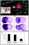
In vivo schematic for therapeutic paradigm of CD-NSPCs and brain metastatic melanoma. A. Eight days after intracarotid injection of 100,000 B16/F10 melanoma cells and establishment of brain metastases, CD-expressing NSPCs (CD-NSPCs) were injected intracranially at rostral and caudal cortical sites. Three days after introduction of CD-NSPCs, mice were treated for eight days with daily i.p. injections of 5-FC. B. Schematic of bystander effect. NSPCs expressing CD migrate to tumor areas, and in the presence of systemically administered pro-drug 5-FC, convert it to 5-FU. The 5-FU and its toxic metabolites diffuse out of the stem cells to selectively kill surrounding, dividing tumor cells. C and D. Tumor areas (outlined in red) in brains of mice that had received injections of melanoma cells only (C) and in brains of mice that had received melanoma cells and were subsequently treated with CD-NSPCs and 5-FC (D). The tumor region has expanded and occupied the majority of the left hemisphere in mice that had received injections of melanoma cells only (two representative animal brain sections shown in panel C). Mice that had received melanoma cells followed by injections of CD-NSPCs and treatment with 5-FC showed a marked decrease in tumor burden (two representative mouse brain sections shown in panel D). C and D. Scale bar, 5 mm. E. Quantitative analysis of tumor volume showing 71% and 69% less tumor burden in mice that had been treated with CD-NSPCs and 5-FC when compared to control group 1 (tumor only, no treatment) or control group 2 (tumor + NSPCs + 5-FC), respectively (mean ± SD; Student’s t-test; n = 4 in each group). Tumor volumes were expressed as percent of control group 1 tumor (no treatment was taken as 100%). All mice were sacrificed 19 days after initiation of melanoma growth by injection of melanoma cells into the left internal carotid artery.

References

    1. Aboody KS, Brown A, Rainov NG, Bower KA, Liu S, Yang W, Small JE, Herrlinger U, Ourednik V, Black PM, Breakefield XO, Snyder EY. Neural stem cells display extensive tropism for pathology in adult brain: Evidence from intracranial gliomas (erratum in Proc. Natl. Acad. Sci. USA [2001] 98, 777). . Proc Natl Acad Sci USA. 2000;97:12846–12851. - PMC - PubMed
    1. Bagnato A, Rosano L, Spinella F, Di Castro V, Tecce R, Natali PG. Endothelin B receptor blockade inhibits dynamics of cell interactions and communications in melanoma cell progression. Cancer Res. 2004;64:1436–1443. - PubMed
    1. Barresi V, Belluardo N, Sipione S, Mudo G, Cattaneo E, Condorelli DF. Transplantation of prodrug-converting neural progenitor cells for brain tumor therapy. Cancer Gene Ther. 2003;10:396–402. - PubMed
    1. Benedetti S, Pirola B, Pollo B, Magrassi L, Bruzzone MG, Rigamonti D, Galli R, Selleri S, Di Meco F, De Fraja C, Vescovi A, Cattaneo E, Finocchiaro G. Gene therapy of experimental brain tumors using neural progenitor cells. Nat Med. 2000;6:447–450. - PubMed
    1. Brown AB, Yang W, Schmidt NO, Carroll R, Leishear KK, Rainov NG, Black PM, Breakefield XO, Aboody KS. Intravascular delivery of neural stem cell lines to target intracranial and extracranial tumors of neural and non-neural origin. Hum Gene Ther. 2003;14:1777–1785. - PubMed

Publication types