Comparative Study
. 2006 Mar 8;26(10):2684-91.
doi: 10.1523/JNEUROSCI.5257-05.2006.

Novel subcellular distribution pattern of A-type K+ channels on neuronal surface

Affiliations
Comparative Study

Novel subcellular distribution pattern of A-type K+ channels on neuronal surface

Mihaly Kollo et al. J Neurosci. .

Abstract

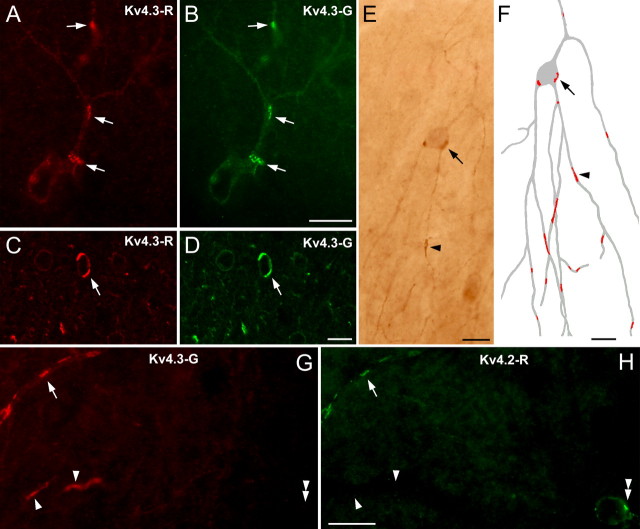
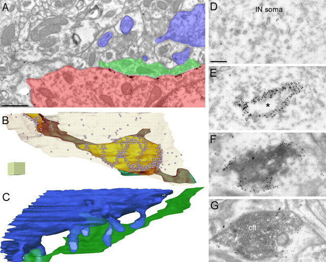
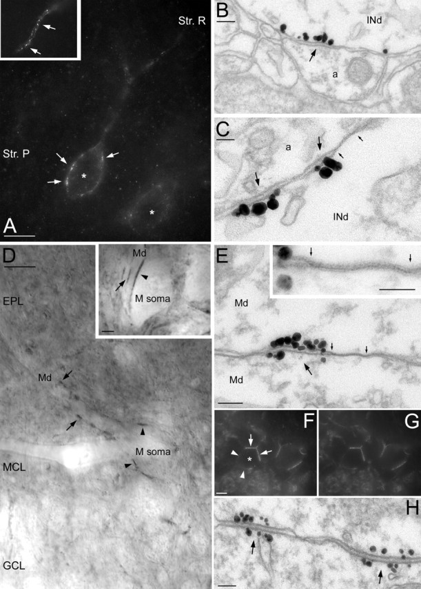
Potassium channels comprise the most diverse family of ion channels. In nerve cells, their critical roles in synaptic integration and output generation have been demonstrated. Here, we provide evidence for a distribution that predicts a novel role of K+ channels in the CNS. Our experiments revealed a highly selective clustering of the Kv4.3 A-type K+ channel subunits at specialized junctions between climbing fibers and cerebellar GABAergic interneurons. High-resolution ultrastructural and immunohistochemical experiments demonstrated that these junctions are distinct from known chemical and electrical (gap junctions) synapses and also from puncta adherentia. Each cerebellar interneuron contains many such K+ channel-rich specializations, which seem to be distributed throughout the somatodendritic surface. We also show that such K+ channel-rich specializations are not only present in the cerebellum but are widespread in the rat CNS. For example, mitral cells of the main olfactory bulb establish Kv4.2 subunit-positive specializations with each other. At these specializations, both apposing membranes have a high density of K+ channels, indicating bidirectional signaling. Similar specializations with pronounced coclustering of the Kv4.2 and 4.3 subunits were observed between nerve cells in the medial nucleus of the habenula. Based on our results and on the known properties of A-type K+ channels, we propose that strategically clustered K+ channels at unique membrane specializations could mediate a novel type of communication between nerve cells.


Figures

Figure 1.
Figure 1.
Clustered distribution of A-type potassium channels in the cerebellum. A, B, Light microscopic images of the cerebellar cortex demonstrate the uneven distribution of Kv4.3 subunits in molecular layer INs (clusters indicated by arrows). Identical labeling was obtained with two antibodies against the Kv4.3 subunit (Kv4.3-R, A, C; Kv4.3-G, B, D), indicating the specificity of the immunoreaction. C, D, Prominent clustering of the immunosignal (e.g., arrows) is shown in confocal laser-scanning microscopic images. E, The nonuniform distribution of the Kv4.3 subunit is also observed in peroxidase reactions, a high-sensitivity technique that allows LM reconstruction of immunostained INs. F, The reconstruction illustrates that a single IN (gray) contains several dozens of clusters (red) at somatic (arrow) and dendritic (arrowhead) locations. G, H, Double immunofluorescent labeling for Kv4.3 (G) and Kv4.2 (H) subunits shows the coclustering (e.g., arrow) of these subunits in some IN dendrites. Several Kv4.3-positive clusters do not contain the Kv4.2 subunit (arrowheads), whereas few Kv4.2 immunopositive clusters (double arrowhead) are immunonegative for Kv4.3. Scale bars, 10 μm.
Figure 2.
Figure 2.
Electron microscopic demonstration of the clustering of the Kv4.3 subunit in specialized junctions. A, B, Part of the dendritic plasma membrane of a cerebellar IN (INd), which is in direct contact with an axon terminal (cft), contains a large number of gold particles (arrows). The axon terminal (cft) establishes three asymmetrical synapses (arrowheads) with PC spines (s) and has ultrastructural features of a CF terminal. The micrograph in B illustrates the consistency of the labeling in a serial section. By tilting the section in the EM, the rigid appositions between the membranes (arrows) are better seen. C, Higher-magnification view of Kv4.3 immunopositive clusters (arrows) on an IN soma. The CF terminal (cft) establishes an asymmetrical synapse (arrowhead) on a spine (s). D, Nonuniform Kv4.3 immunoreactivity is shown in an IN dendrite (INd). Gold particles are clustered in the dendritic membrane (arrows), where it is in contact with either the CF terminal (cft) or with its preterminal axon (pta). E, Double fluorescent labeling for Kv4.3 (green) and vGluT2 (red) demonstrates that Kv4.3 clusters are apposed to vGluT2 immunopositive CF terminals. The inset shows the lack of full overlap of the green and red spots. F, EM double labeling for Kv4.3 (particles) and vGluT2 (peroxidase reaction), in which the preterminal axon (pta) of a vGluT2-positive CF contacts an IN dendrite (INd) clustering of Kv4.3 (arrow) is detected. Note that the DAB reaction is present in the CF axon terminal (cft) but not in the preterminal axon. Scale bars: A–D, 0.2 μm; E, 15 μm; E, inset, 2 μm; F, 0.4 μm.
Figure 3.
Figure 3.
Complex structures of the Kv4.3 subunit immunopositive specializations. A, Electron micrograph of the cerebellar molecular layer showing clusters of Kv4.3 immunogold particles along the somatic plasma membrane of an IN (red). The CF terminal is green, and the PC dendrite and spines are blue. The illustrated micrograph is one of the 44 serial sections on which the 3D reconstruction (shown in B and C) is based. B, Three-dimensional reconstruction of a CF terminal (green; brown when viewed through the transparent IN membrane) viewed through the transparent IN somatic membrane (light cream). The area of the IN plasma membrane, which is in direct contact with the CF, is yellow. Gold particles are illustrated with small spheres. Note that the high concentration of gold particles does not cover the entire yellow area, but a complex mosaic of K+ channel-rich domains can be seen. C, The same 3D reconstruction illustrating the Purkinje dendrite and spines (blue) and the CF terminal. Several excitatory synapses (light blue) on PC spines (blue) are established by the CF terminal (green), viewed from the opposite side as in B. D–G, Electron micrographs of consecutive serial sections of an IN, demonstrating the nonhomogeneous labeling of the membrane apposition between a vGluT2-positive CF terminal (peroxidase reaction) and an IN soma. En face view of the membrane apposition, showing a strongly immunopositive ring with a negative center (*). Scale bars: A, 0.8 μm; B, C, each side of the cube is 0.5 μm; D–G, 0.2 μm.
Figure 4.
Figure 4.
Structural evidence that the Kv4.3 subunit-rich specializations are distinct from known chemical synapses. A, A CF terminal in the cerebellar molecular layer establishes an asymmetrical (type I) chemical synapse (arrow) with a PC spine (s) and several membrane specializations (arrowheads) with an IN soma. B–D, High-magnification views of a membrane specialization between a CF terminal and an IN (B), an asymmetrical (type I) chemical synapse made on a Purkinje spine (C), a symmetrical (type II) synaptic junction (D, right), and a punctum adherens (D, left) on an IN dendrite. The presence of presynaptic vesicle clustering distinguishes type I and type II synapses from the CF-IN membrane specializations. Rigid apposition of the presynaptic and postsynaptic membranes characterizes all types of junctions. Scale bars: A, 100 nm; B–D, 50 nm (same magnification).
Figure 5.
Figure 5.
Immunohistochemical demonstration that the membrane specializations between CFs and INs are distinct from glutamatergic synapses. A, Postembedding immunogold localization of the GluR2/3 glutamate receptor subunits in the cerebellar molecular layer. Gold particles are concentrated in the asymmetrical synapses (arrows) made by a CF terminal (cft) with Purkinje cell spines (s), but the membrane apposition between the terminal and the IN somatic plasma membrane (arrowheads) is immunonegative. B, C, Postembedding immunogold localization of the NMDA receptor NR2A/B subunits in the cerebellum (B) and in the hippocampus (C). The membrane specialization between a CF terminal and an IN soma (B, arrowheads) does not contain any gold particle for the NR2A/B subunits, but in the same reactions, Schaffer collateral synapses (C, arrows) on hippocampal CA1 pyramidal cell spines (s) are strongly immunopositive. D, Pre-embedding immunogold reaction for mGluR1α in the cerebellar molecular layer. The membrane apposition (arrowheads) between a CF terminal (cft) and an IN dendrite contains gold particles at approximately the same density as in the rest of the IN plasma membrane. Scale bars, 0.2 μm.
Figure 6.
Figure 6.
A-type K+ channel-rich specializations are present throughout the CNS. A, LM immunofluorescent labeling for the Kv4.3 subunit in the hippocampal CA1 area. Kv4.3 immunopositive interneurons (*) are shown with several intensely labeled clusters (arrows) decorating the somatic plasma membrane. The inset illustrates that dendritic immunoreactivity is also highly nonuniform. B, C, EM demonstration of the clustering of gold particles (arrows) for Kv4.3 subunit in hippocampal IN dendrites (INd). A gap junction (marked by small arrows in C) is found next to the K+ channel-rich specialization. D, Nonuniform distribution of the Kv4.2 subunit in mitral cells of the main olfactory bulb. Several intensely labeled spots are present in the somatic (arrowheads in M soma) and dendritic (arrows in Md) membranes of a mitral cell. The inset shows strongly immunopositive clusters between a mitral cell soma and a dendrite (arrowhead) and between two dendrites (arrow). E, Clustering of gold particles (arrow) between two mitral cell dendrites (Md). A large number of immunogold particles for the Kv4.2 subunits is present at both sides of the specialization. Note the presence of a gap junction (marked by small arrows) next to the gold cluster. The gap junction is illustrated at a higher magnification in the inset. F, G, Double immunofluorescent labeling for Kv4.3 (F) and Kv4.2 (G) subunits in the medial habenular nucleus. The somata of immunopositive cells (*) are not uniformly labeled, but strongly immunopositive clusters (arrows) are found where cell bodies are in direct contact with each other, whereas the rest of the somatic membrane (arrowheads) is only weakly labeled. H, EM immunogold localization of the Kv4.2 subunit in the medial habenula demonstrates the clustering of particles at both membranes forming the specializations (arrows). Str. P, Stratum pyramidale; Str. R, stratum radiatum; EPL, external plexiform layer; GCL, granule cell layer; MCL, mitral cell layer. Scale bars: A, D, F, G, 10 μm; D, inset, 2 μm; B, E, H, 100 nm; C, E, inset, 50 nm.

References

    1. Alonso G, Widmer H (1997). Clustering of Kv4.2 potassium channels in postsynaptic membrane of rat supraoptic neurons: an ultrastructural study. Neuroscience 77:617–621. - PubMed
    1. An WF, Bowlby MR, Betty M, Cao J, Ling HP, Mendoza G, Hinson JW, Mattsson KI, Strassle BW, Trimmer JS, Rhodes KJ (2000). Modulation of A-type potassium channels by a family of calcium sensors. Nature 403:553–556. - PubMed
    1. Cai X, Liang CW, Muralidharan S, Kao JP, Tang CM, Thompson SM (2004). Unique roles of SK and Kv4.2 potassium channels in dendritic integration. Neuron 44:351–364. - PubMed
    1. Chandy KG (1991). Simplified gene nomenclature. Nature 352:26. - PubMed
    1. Csiffary A, Gorcs TJ, Palkovits M (1990). Neuropeptide Y innervation of ACTH-immunoreactive neurons in the arcuate nucleus of rats: a correlated light and electron microscopic double immunolabeling study. Brain Res 506:215–222. - PubMed

Publication types

MeSH terms

Substances

LinkOut - more resources