Comparative Study
. 2006 Mar 8;26(10):2757-66.
doi: 10.1523/JNEUROSCI.3808-05.2006.

Depolarization of cochlear outer hair cells evokes active hair bundle motion by two mechanisms

Affiliations
Comparative Study

Depolarization of cochlear outer hair cells evokes active hair bundle motion by two mechanisms

Helen J Kennedy et al. J Neurosci. .

Abstract

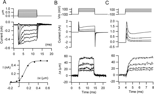
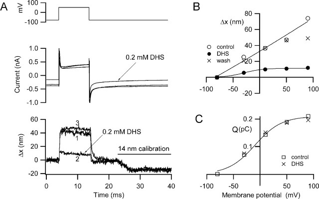
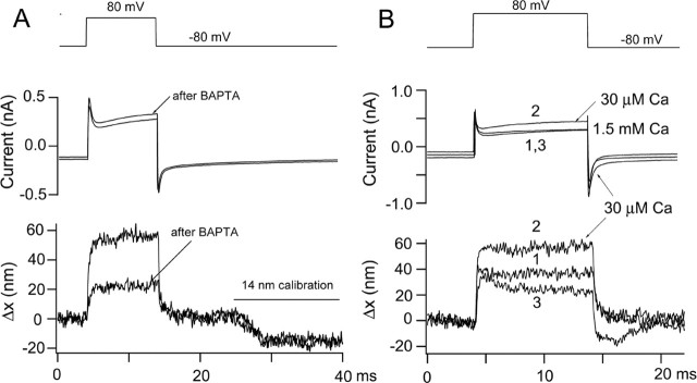
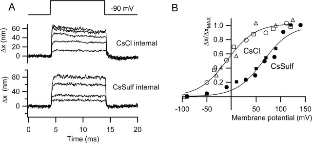
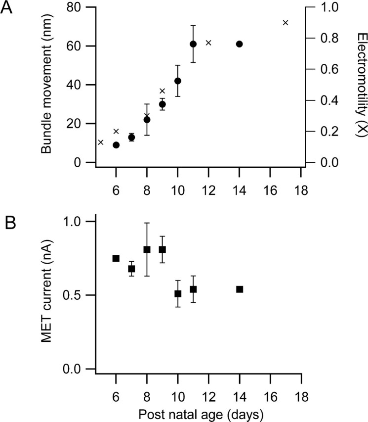
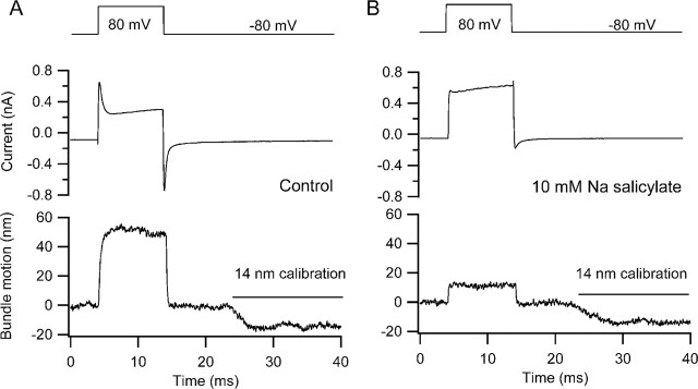
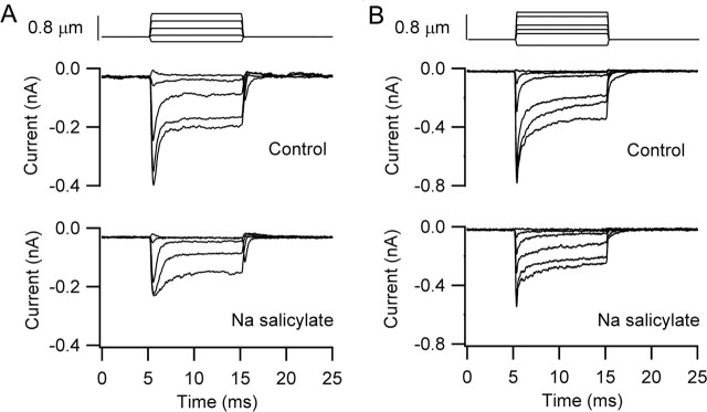
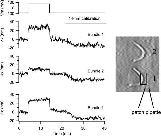
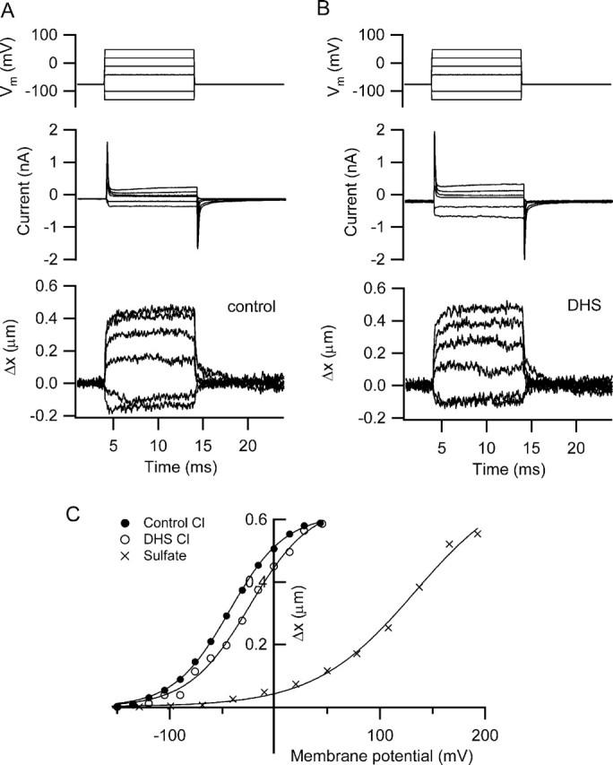
There is current debate about the origin of mechanical amplification whereby outer hair cells generate force to augment the sensitivity and frequency selectivity of the mammalian cochlea. To distinguish contributions to force production from the mechanotransducer (MET) channels and somatic motility, we have measured hair bundle motion during depolarization of individual outer hair cells in isolated rat cochleas. Depolarization evoked rapid positive bundle deflections that were reduced by perfusion with the MET channel blocker dihydrostreptomycin, with no effect on the nonlinear capacitance that is a manifestation of prestin-driven somatic motility. However, the movements were also diminished by Na salicylate and depended on the intracellular anion, properties implying involvement of the prestin motor. Furthermore, depolarization of one outer hair cell caused motion of neighboring hair bundles, indicating overall motion of the reticular lamina. Depolarization of solitary outer hair cells caused cell-length changes whose voltage-activation range depended on the intracellular anion but were insensitive to dihydrostreptomycin. These results imply that both the MET channels and the somatic motor participate in hair bundle motion evoked by depolarization. It is conceivable that the two processes can interact, a signal from the MET channels being capable of modulating the activity of the prestin motor.


Figures

Figure 1.
Figure 1.
Mechanotransducer currents and hair bundle movements in an outer hair cell. A, Imposed deflections of the hair bundle elicit MET currents with pronounced fast adaptation, which has a time constant for small responses, 0.28 ms. The relationship between peak current and bundle displacement is shown (filled circles), which has been fit with a single Boltzmann equation: I = Imax/(1 + exp ((V0.5V)/Vϵ))−1, with Imax of 0.5 nA, V0.5 of 0.11 μm, and Vϵ of 0.053 μm. Holding potential, −84 mV. B, Membrane currents and hair bundle movements in the excitatory direction (toward the tip of the V) for depolarizing voltage steps from a holding potential of −90 mV. Note the large-capacity transients indicative of the charge movements associated with prestin activation. C, Onset of currents and movements shown on a faster timescale. Superimposed on the records are exponential fits with time constants of 0.14, 0.14, 0.16, and 0.16 ms for the decay of the capacity transient for increasing voltage step and 0.26, 0.26, 0.25, and 0.39 ms for onset of the movement. P10 rat, Cs2SO4 intracellular solution.
Figure 2.
Figure 2.
Depolarization-induced hair bundle motion and its block by dihydrostreptomycin. A, Membrane currents (middle) and hair bundle movements, Δx, (bottom) for depolarization to +50 mV before (1), during (2), and after (3) recovery from application of 0.2 mm DHS. DHS reduces the maximum movement and also blocks part of the current turned on at the holding potential (−84 mV). For all Δx traces, movement toward the tip of the V is denoted as positive, and the negative deflections at the end of the traces is a 14 nm calibration signal. B, Voltage dependence of the movements (Δx) before, during, and after DHS application. C, Voltage dependence of charge movement (Q) before and during DHS perfusion. The charge movement (Q), calculated from the current transients at the onset of the depolarizing step, reflects activation of the prestin motor and is unaffected by DHS. Bathing solution contains 0.1 mm Ca2+ and 1 mm Mg2+. P9 rat, CsCl intracellular, maximum MET current of 0.77 nA.
Figure 3.
Figure 3.
Effects of Ca2+ on hair bundle motion. A, Membrane currents and bundle movements for depolarizing voltage steps to +80 mV before and immediately after pressure ejection of submicromolar Ca2+ buffered with 5 mm BAPTA. The manipulation produced a two-thirds decrease in the motion without effect on the capacity transients. The movements have not been scaled for the size of the calibration response, and the change in photodiode current for the 14 nm calibration was virtually identical before and after BAPTA, suggesting no drift or change in the bundle image after BAPTA application. P10 rat, Cs2SO4 intracellular, maximum MET current of 0.35 nA. The MET current had declined to <0.1 nA after the treatment. B, Membrane currents and bundle movements for depolarization to +80 mV before (1), during (2), and after (3) lowering the extracellular Ca2+ concentration from 1.5 mm to 30 μm. Note that Ca2+ reduction increases the sizes of both movement and current. P9 rat, Cs2SO4 intracellular, maximum MET current of 0.71 nA.
Figure 4.
Figure 4.
Effects of intracellular anion on hair bundle motion. A, Families of bundle movements (Δx) recorded in two outer hair cells with different intracellular solutions, CsCl (top) and Cs2SO4 (bottom), for a range of membrane potentials from −90 to +110 mV (CsCl) and −90 to +140 mV (Cs2SO4). Timing of voltage pulse is shown above. B, Plots of bundle movements (Δx) normalized to the maximum motion (Δxmax) versus membrane potential for three cells with CsCl intracellular (open symbols) and two cells with Cs2SO4 intracellular (filled symbols). Note that the steepness of the voltage dependence is similar, but the results in Cs2SO4 are shifted ∼75 mV positive relative to those with CsCl. Smooth curves are fits to the following: Δxxmax = (1 + exp ((V0.5V)/Vϵ))−1, where V0.5 and Vϵ were −9 and 29 mV, respectively, for chloride and +65 and 28 mV for sulfate.
Figure 5.
Figure 5.
Effects of postnatal development on bundle movements and MET currents. A, Maximum hair bundle motion for depolarization to +80 mV increases with postnatal age of the rat (circles). Superimposed on the results is the development of somatic electromotility with postnatal age of rat (crosses: normalized data taken from Belyantseva et al., 2000, their Fig. 4) referred to the right axis. Note that, according to their normalization, maximum somatic motility is not achieved until after P20 and, at P17, is 0.9 of the adult. B, Peak mechanotransducer currents at −80 mV holding potential changes little over the same developmental period. Each point is the mean ± SEM of two to nine measurements. Both measurements at P14 were from hearing animals.
Figure 6.
Figure 6.
Effects of Na salicylate on hair bundle movements. A, Membrane current and bundle movement for depolarization to +80 mV before applying Na salicylate. B, Membrane current and bundle movement for depolarization to +80 mV 9 min after starting perfusion with 10 mm Na salicylate. No recovery was obtained on return to control solution. Na salicylate blocks the somatic motor, as indicated by the disappearance of the nonlinear capacitance reflected by the current transients at the onset and termination of the voltage step. P11 rat, Cs2SO4 intracellular, maximum MET current of 0.40 nA.
Figure 7.
Figure 7.
Effects of Na salicylate on mechanotransducer currents. A, MET currents before and 15 min after 10 mm Na salicylate perfusion for the cell of Figure 6. The test MET currents were acquired after the records in Figure 6B when the nonlinear capacitance in response to depolarizing voltage steps had disappeared. P11 rat, Cs2SO4 intracellular, maximum MET current of 0.40 nA. B, MET currents before and 30 min after 10 mm Na salicylate perfusion. The test MET currents were acquired once the nonlinear capacitance had disappeared (data not shown). P7 rat, CsCl intracellular, maximum MET current of 0.73 nA.
Figure 8.
Figure 8.
Depolarization evokes motion of adjacent hair bundle. Bundle movements when the photodiode was positioned on the bundle of the cell being depolarized (trace 1, bundle 1). Photodiode was positioned on the bundle of the next cell along in the row (trace 2, bundle 2). On returning the photodiode to bundle 1, (trace 3), the same size of response was elicited. For each movement record, the negative excursion at the end of the trace is the 14 nm calibration. On the right is shown two hair bundles, the location of the patch pipette, and the photodiode pair superimposed on bundle 1. P10 rat, CsCl intracellular, maximum MET current of 1 nA.
Figure 9.
Figure 9.
Somatic contractions measured in rat isolated hair cells. A, Membrane currents (middle traces) and changes in length (Δx, bottom traces) in an outer hair cell in response to a series of depolarizing and hyperpolarizing voltage steps. Intracellular solution contained CsCl. B, Same measurements made 5 min after starting perfusion with 0.2 mm DHS, indicating no effect of the antibiotic on maximum movements or modest effect on the currents. C, Dependence of length change, Δx, on membrane potential for the responses in A (filled circles) and B (open circles). Also shown is the variation of length change with membrane potential in another outer hair cell recorded with an intracellular solution containing Cs2SO4 and gluconate as the principal external anion. Smooth curves are fits to the following: Δxxmax = (1 + exp ((V0.5V)/Vϵ))−1, where V0.5 and Vϵ were −43 and 27 mV, respectively, for control chloride, −26 and 29 mV in the presence of DHS, and +133 and 48 mV for sulfate. Outer hair cells isolated from a P11 rat (DHS experiment) and a P13 rat (Cs2SO4 experiment).

Similar articles

Cited by

References

    1. Ashmore JF (1987). A fast motile response in guinea-pig outer hair cells: the cellular basis of the cochlear amplifier. J Physiol (Lond) 388:323–347. - PMC - PubMed
    1. Ashmore JF (1989). Transducer motor coupling in cochlea outer hair cells. In: Cochlear mechanisms: structure, function and models (Wilson JP, Kemp DT, eds) , pp. 107–114. New York: Plenum.
    1. Assad JA, Hacohen N, Corey DP (1989). Voltage dependence of adaptation and active bundle movement in bullfrog saccular hair cells. Proc Natl Acad Sci USA 86:2918–2922. - PMC - PubMed
    1. Assad JA, Shepherd GM, Corey DP (1991). Tip-link integrity and mechanical transduction in vertebrate hair cells. Neuron 7:985–994. - PubMed
    1. Belyantseva IA, Adler HJ, Curi R, Frolenkov GI, Kachar B (2000). Expression and localization of prestin and the sugar transporter GLUT-5 during development of electromotility in cochlear outer hair cells. J Neurosci 20: RC116 (1–5). - PMC - PubMed

Publication types

MeSH terms

LinkOut - more resources