Comparative Study
. 2006 Mar 8;26(10):2777-87.
doi: 10.1523/JNEUROSCI.3420-05.2006.

Glial cell line-derived neurotrophic factor-dependent recruitment of Ret into lipid rafts enhances signaling by partitioning Ret from proteasome-dependent degradation

Affiliations
Comparative Study

Glial cell line-derived neurotrophic factor-dependent recruitment of Ret into lipid rafts enhances signaling by partitioning Ret from proteasome-dependent degradation

Brian A Pierchala et al. J Neurosci. .

Abstract

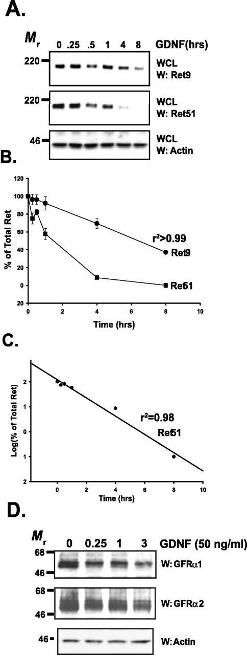
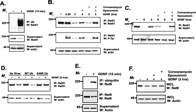
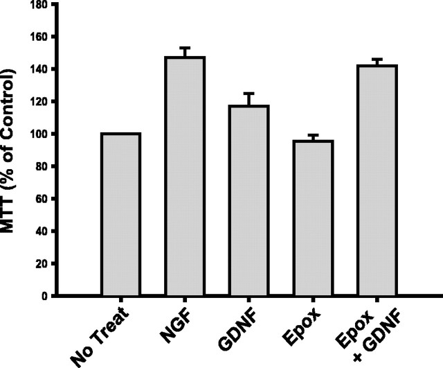
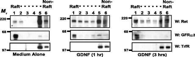
The receptor tyrosine kinase (RTK) Ret is activated by the formation of a complex consisting of ligands such as glial cell line-derived neurotrophic factor (GDNF) and glycerophosphatidylinositol-anchored coreceptors termed GFRalphas. During activation, Ret translocates into lipid rafts, which is critical for functional responses to GDNF. We found that Ret was rapidly ubiquitinated and degraded in sympathetic neurons when activated with GDNF, but, unlike other RTKs that are trafficked to lysosomes for degradation, Ret was degraded predominantly by the proteasome. After GDNF stimulation, the majority of ubiquitinated Ret was located outside of lipid rafts and Ret was lost predominantly from nonraft membrane domains. Consistent with the predominance of Ret degradation outside of rafts, disruption of lipid rafts in neurons did not alter either the GDNF-dependent ubiquitination or degradation of Ret. GDNF-mediated survival of sympathetic neurons was inhibited by lipid raft depletion, and this inhibitory effect of raft disruption on GDNF-mediated survival was reversed if Ret degradation was blocked via proteasome inhibition. Therefore, lipid rafts sequester Ret away from the degradation machinery located in nonraft membrane domains, such as Cbl family E3 ligases, thereby sustaining Ret signaling.


Figures

Figure 1.
Figure 1.
Ret and GFRαs are degraded rapidly after activation. A, Sympathetic neurons were treated with GDNF (50 ng/ml) for various lengths of time, whole-cell extracts were produced from them, and the extracts were subjected to immunoblotting. Ret9 (top) and Ret51 (middle) immunoblotting revealed that both of these Ret isoforms are lost rapidly after GDNF stimulation. Actin immunoblotting (bottom) was used to confirm equal loading of cell extracts. WCL, Whole-cell lysate. B, Quantification of the experiment shown in A. Three separate experiments were conducted, quantified, and graphed as the mean ± SEM. C, Semilog plot of the data for Ret51 degradation kinetics shown in B. D, Sympathetic neurons were treated with GDNF (50 ng/ml) for various lengths of time (labeled on top), and whole-cell extracts were prepared. These extracts were immunoblotted with antibodies to GFRα1 (top) and GFRα2 (middle). Equal loading of proteins was confirmed with actin immunoblotting (bottom). This experiment was conducted twice with identical results. W, Western blot.
Figure 2.
Figure 2.
Ret is ubiquitinated and degraded predominantly by the proteasome. A, Sympathetic neurons were treated with GDNF (50 ng/ml) or medium alone for 15 min, lysed, and ubiquitinated proteins were immunoprecipitated. The immunoprecipitates were analyzed by Ret51 immunoblotting (top). The supernatants from the immunoprecipitation were subjected to Ret51 and actin immunoblotting to determine the amount of nonubiquitinated Ret51 and to confirm that equal amounts of protein were analyzed, respectively. B, Neurons were pretreated with epoxomicin (5 μm), concanamycin (10 μm), or both for 30–60 min before stimulation with GDNF for 3 h. As a control, some neurons were pretreated with medium containing the vehicle alone (DMSO) for 1 h. After the treatments, the neurons were detergent extracted, and Ret51 was immunoprecipitated. These immunoprecipitates were subjected to Ret51 immunoblotting (top). The blots were stripped and reprobed with phosphotyrosine antibodies (P-Tyr; middle) and the supernatants of the immunoprecipitates analyzed with actin immunoblotting (bottom) to confirm that similar amounts of protein were analyzed. C, Sympathetic neurons were treated with GDNF, epoxomicin, or concanamycin for 8 h, similar to the analysis in B. Ret51 analysis was performed as described in B. D, Neurons were either not infected (No Virus) or infected with lentivirus to express either wild-type (WT) ubiquitin or ubiquitin containing a K48R mutation for 4 d. The medium was then replaced, and 24 h later, these neurons were treated with GDNF (50 ng/ml) for 2 h. The levels of Ret51 were then examined as described in B. E, Neurons that were treated with GDNF (50 ng/ml) for 15 min were subjected to ubiquitin immunoprecipitation, as in A. These precipitates were immunoblotted for Ret9 (top), and the supernatants were subjected to Ret9 (middle) and actin (bottom) immunoblotting. F, Sympathetic neurons were subjected to an identical analysis as in B. Whole-cell lysates (WCLs) were made from the treated neurons, and these were analyzed by Ret9 (top) and actin (bottom) immunoblotting. All of the experiments described in this figure were performed two to four times with identical results. IP, Immunoprecipitate; Ub, ubiquitin; W, Western blot.
Figure 3.
Figure 3.
Ligand-dependent Ret activation limits the trophic activity of GDNF on sympathetic neurons. The metabolic activity of neurons was determined using an MTT reduction assay as described in Materials and Methods. Neurons were treated with NGF (50 ng/ml), GDNF (50 ng/ml), epoxomicin (Epox; 5 μm), or GDNF in the presence of epoxomicin for 4 h. MTT reduction was then measured and graphed as a percentage of neurons treated with medium alone (No Treat). Error bars represent SEM.
Figure 4.
Figure 4.
Ret translocates into lipid rafts after activation with GDNF. Sympathetic neurons were treated with medium alone or with GDNF (50 ng/ml) for either 1 or 3 h. The neurons were then lysed and subjected to Optiprep density centrifugation as described in Materials and Methods. Six equal fractions were taken from the top of the tubes and labeled from 1 to 6, fraction 6 being the densest (raft and nonraft fractions are labeled above the fraction numbers). These fractions were analyzed by immunoblotting for Ret51 (top) and GFRα2 (middle). GFRα2 confirmed the location of the buoyant, lipid raft fractions, and immunoblotting with antibodies to transferrin receptor (TrfR; bottom) indicated the nonraft membrane fractions. This experiment was performed twice with similar results. W, Western blot.
Figure 5.
Figure 5.
Ubiquitinated Ret resides outside of lipid rafts in GDNF-stimulated sympathetic neurons. A, Sympathetic neurons were stimulated with GDNF (50 ng/ml) for various lengths of time, and the neurons were then extracted with Triton X-100. The extracts were centrifuged, and the insoluble pellets containing lipid rafts (left 4 lanes) and the soluble fraction containing nonraft membrane fractions (right 4 lanes) were then separated by SDS-PAGE and electroblotted. The blots were probed with antibodies to Ret9 (top 2 panels), Ret51 (next 2 panels), and PY1062Ret (5th panel). Shorter exposures of the Ret9 and Ret51 immunoblots are shown (labeled as “Light,” panels 2 and 4) to better observe the loss of Ret protein in these fractions. To confirm the relative purity of the lipid-raft fractionation, blots were probed with anti-transferrin receptor (6th panel) and anti-caveolin-1 (bottom panel), which are markers of nonraft fractions and lipid-raft fractions, respectively. B, Sympathetic neurons were treated with GDNF (50 ng/ml) for 15 min and then Triton X-100 was extracted as described in A. The fractions were then treated with glucoside to solubilize the rafts and subjected to ubiquitin immunoprecipitation. The supernatants of this immunoprecipitation were used for a second Ret51 immunoprecipitation, and all of the immunoprecipitates were subjected to Ret51 immunoblotting (top). The activation state of the complexes was determined with phosphotyrosine immunoblotting (second), and the purity of the raft and nonraft fractions was confirmed by probing the supernatants with antibodies to transferrin receptor and caveolin-1 (bottom 2 panels). These immunoblots also confirmed that equal amounts of protein were analyzed. The experiments in this figure were conducted two or three times with similar results. INSOL, Insoluble pellets containing lipid rafts; SOL, soluble fraction containing nonraft membrane fractions; IP, immunoprecipitate; TrfR, transferrin receptor; Cav, caveolin; Ub, ubiquitin; P-Tyr, phosphotyrosine; W, Western blot.
Figure 6.
Figure 6.
Lipid rafts are not required for the GDNF-induced ubiquitination or degradation of Ret. A, Sympathetic neurons were treated with MCD (10 mm), or medium alone, for 15 min, washed, and then stimulated with GDNF (50 ng/ml) or medium alone for 1 h. The neurons were extracted with 1% Triton X-100, and the insoluble pellets (left) containing lipid rafts were collected via centrifugation from the soluble, nonraft fractions (right). These fractions were separated by SDS-PAGE and immunoblotted with antibodies to Ret51. To confirm the relative purity of the insoluble and soluble fractions, flotillin and transferrin receptor immunoblotting, respectively, was conducted. B, Neurons were treated with MCD as in A, washed, and then stimulated with GDNF (50 ng/ml), or medium alone, for 3 h. Whole-cell extracts were then produced from the neurons, and the extracts were subjected to Ret9 (top) and Ret51 (2nd panel) immunoblotting. Actin immunoblotting confirmed that equal amounts of protein were analyzed. C, Sympathetic neurons were treated with medium alone or with neutral Smase (2 mU/ml) for 90 min. The neurons were then stimulated with GDNF (50 ng/ml) or vehicle alone for 1 h in the continued presence of Smase. These neurons were then subjected to Triton X-100 extraction and immunoblotting as described in A. D, Similar to the treatments described in C, sympathetic neurons were treated with Smase before treatment with GDNF or medium alone for 3 h. Total cellular protein was harvested, and analyzed as was done as in B. E, Cells were treated with MCD, Smase, or vehicle only, as done in the previous panels. After a 15 min treatment with GDNF (50 ng/ml), the neurons were detergent extracted, and ubiquitinated proteins were immunoprecipitated. These immune complexes were analyzed with Ret51 immunoblotting, and actin immunoblotting of the supernatants confirmed that equal amounts of protein were analyzed. The experiments in this figure were performed two to four times with similar results. INSOL, Insoluble pellets containing lipid rafts; SOL, soluble fraction containing nonraft membrane fractions; IP, immunoprecipitate; TrfR, transferrin receptor; Cav, caveolin; W, Western blot; WCL, whole-cell lysate.
Figure 7.
Figure 7.
Lipid rafts enhance the survival-promoting activity of GDNF by protecting Ret from degradation. Sympathetic neurons were maintained in NGF, deprived of NGF for 24 h, or deprived of NGF in the presence of GDNF (50 ng/ml). Some neurons that were treated with GDNF were concurrently treated with the proteasome inhibitor epoxomicin (Epox; 5 μm) or were depleted of cholesterol by treatment with lovastatin (Lova; 5 μm) for 12 h before GDNF treatment in the continued presence of the inhibitor. As a control, NGF-deprived neurons were saved with potassium depolarization alone (KCl; 25 mm) or in the presence of lovastatin. The neurons were then fixed, Nissl stained, and the number of surviving neurons in each condition counted. These experiments were plotted as the mean ± SEM. The asterisks indicate statistically significant differences (p < 0.05) from the two indicated conditions and GDNF or GDNF and lovastatin treatments, respectively.
Figure 8.
Figure 8.
Lipid rafts protect Ret from degradation by sequestering Ret away from Cbl proteins. Sympathetic neurons were treated with medium alone or with medium containing GDNF (50 ng/ml) for 45 min. Detergent-resistant membranes were purified as described in Materials and Methods. Cbl-b was immunoprecipitated from both Triton X-100-insoluble and -soluble fractions, and these immunoprecipitates were analyzed by immunoblotting using the antibodies listed to the right of each blot. The relative purity of the lipid raft fractions were confirmed with flotillin-1 and transferrin receptor immunoblotting of the supernatants from the immunoprecipitations. This experiment was performed twice with similar results. INSOL, Insoluble pellets containing lipid rafts; SOL, soluble fraction containing nonraft membrane fractions; IP, immunoprecipitate; TrfR, transferrin receptor; P-Tyr, phosphotyrosine; W, Western blot.

Similar articles

Cited by

References

    1. Aguilar RC, Wendland B (2003). Ubiquitin: not just for proteasomes anymore. Curr Opin Cell Biol 15:184–190. - PubMed
    1. Airaksinen MS, Saarma M (2002). The GDNF family: signalling, biological functions and therapeutic value. Nat Rev 3:383–394. - PubMed
    1. Anderson RG (1998). The caveolae membrane system. Annu Rev Biochem 67:199–225. - PubMed
    1. Arenas E, Trupp M, Akerud P, Ibanez CF (1995). GDNF prevents degeneration and promotes the phenotype of brain noradrenergic neurons in vivo. Neuron 15:1465–1473. - PubMed
    1. Baloh RH, Enomoto H, Johnson EM Jr, Milbrandt J (2000). The GDNF family ligands and receptors-implications for neural development. Curr Opin Neurobiol 10:103–110. - PubMed

Publication types

MeSH terms

LinkOut - more resources