Comparative Study
. 2006 Mar 8;26(10):2798-807.
doi: 10.1523/JNEUROSCI.4720-05.2006.

Physiological modulation of intestinal motility by enteric dopaminergic neurons and the D2 receptor: analysis of dopamine receptor expression, location, development, and function in wild-type and knock-out mice

Affiliations
Comparative Study

Physiological modulation of intestinal motility by enteric dopaminergic neurons and the D2 receptor: analysis of dopamine receptor expression, location, development, and function in wild-type and knock-out mice

Zhi Shan Li et al. J Neurosci. .

Abstract

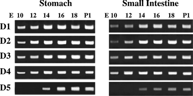
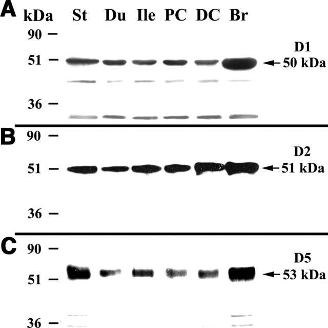
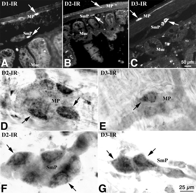
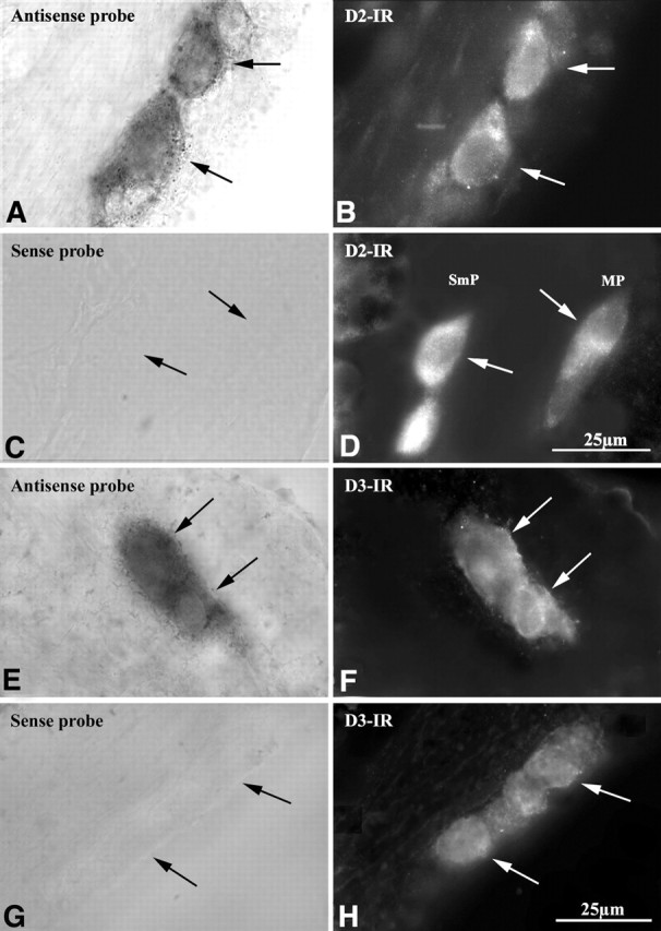
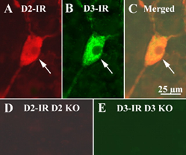
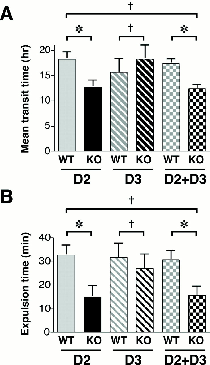
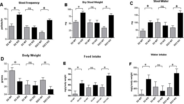
Dopaminergic neurons are present in both plexuses of the murine bowel and are upregulated after extrinsic denervation but play unknown roles in enteric nervous system (ENS) physiology. Transcripts encoding dopamine (DA) receptors D1-D5 were analyzed by reverse transcription-PCR in stomach approximately duodenum approximately ileum approximately proximal > > distal colon. Dissected muscle and myenteric plexus contained transcripts encoding D1-D3 and D5, whereas mucosa contained D1 and D3-D5. D1-D5 expression began in fetal gut [embryonic day 10 (E10)], before the appearance of neurons (E12), and was sustained without developmental regulation through postnatal day 1. In situ hybridization revealed that subsets of submucosal and myenteric neurons contained mRNA encoding D2 or D3. Immunoblots confirmed that D1, D2, and D5 receptor proteins were present from stomach through distal colon. Subsets of submucosal and myenteric neurons were also D1, D2, or D3 immunoreactive. When double labeled by in situ hybridization, these neurons contained mRNA encoding the respective receptors. Total gastrointestinal transit time (TGTT) and colonic transit time (CTT) were measured in mice lacking D2, D3, or D2 plus D3. Both TGTT and CTT were decreased significantly (motility increased) in D2 and D2 plus D3, but not D3, knock-out animals. Mice lacking D2 and D2 plus D3 but not D3 were smaller than wild-type littermates, yet ate significantly more and had greater stool frequency, water content, and mass. Because motility is abnormal when D2 is absent, the net inhibitory DA effect on motility is physiologically significant. The early expression of DA receptors is also consistent with the possibility that DA affects ENS development.


Figures

Figure 1.
Figure 1.
DA receptors are expressed in the gut. Expression of transcripts encoding the dopamine receptors D1–D5 was analyzed regionally in the whole gut wall (A) and in dissected layers of the wall of the ileum (B, C). A, Transcripts encoding each of the five DA receptors were detected in the stomach (St), duodenum (Du), ileum (Ile), proximal colon (PC), and distal (DC) colon. The brain (Br) was investigated as a positive control. B, The presence of transcripts encoding the neural marker β3-tubulin and mucosal epithelial marker sucrase-isomaltase was studied to assess the potential contamination of mucosal preparations with RNA from neurons and LMMP preparations with RNA from the mucosal epithelium. As expected, preparations from Br, whole ileum, and LMMP, but not the mucosa (Muc), contained transcripts encoding β3-tubulin. In contrast, transcripts encoding sucrase-isomaltase were detected in preparations from whole ileum and mucosa but not in those from brain or LMMP. Cross-contamination, therefore, was negligible. M, Molecular marker. C, Transcripts encoding D1, D3, and D5 were present both in the mucosa and the LMMP; those encoding D2 were present in LMMP but not in mucosa; those encoding D4 were present in mucosa but not in LMMP.
Figure 2.
Figure 2.
Expression of DA receptors begins early in fetal development and persists in the postnatal gut. DA receptor expression was analyzed by reverse transcription-PCR in the stomach and small intestine from fetal day E10 through postnatal day P1. Transcripts encoding DA receptors D1–D4 were detected as early as E10 in both the presumptive stomach and small intestine. Transcripts encoding D5 were not detected in either organ until E14. Expression of each receptor was detected through P1.
Figure 3.
Figure 3.
DA receptor immunoreactivity was detected in enteric neurons of mouse ileum. The immunoreactivity was visualized with antibodies to D1, D2, and D3 in both frozen section (A–C) and in whole-mount preparations (D–G). A, D1 immunoreactivity is present in the myenteric (MP) and submucosal (SmP) plexuses and in the mucosa (Muc). The arrows indicate the immunoreactive products. B, D, F, D2 immunoreactivity is present in subsets of myenteric and submucosal neurons, but not in the mucosa. C, E, G, D3 immunoreactivity is present in subsets of myenteric and submucosal neurons. Mucosal immunoreactivity is very weak. Scale bars: (in C) A–C, 5μm; (in G) D–G, 25μm.
Figure 4.
Figure 4.
DA receptor immunoreactivity was detected in enteric neurons of mouse ileum. The immunoreactivity was visualized with antibodies to D1, D2, and D3 in both frozen sections (A–C) and in whole-mount preparations (D–G). A, D1 immunoreactivity is present in the myenteric (MP) and submucosal (SmP) plexuses and in the mucosa (Muc). The arrows indicate the immunoreactive products. B, D, F, D2 immunoreactivity is present in subsets of myenteric and submucosal neurons, but not in the mucosa. C, E, G, D3 immunoreactivity is present in subsets of myenteric and submucosal neurons. Mucosal immunoreactivity is very weak. Scale bars: (in C) A–C, 50 μm; (in G) D–G, 25 μm.
Figure 5.
Figure 5.
Combined in situ hybridization and immunocytochemistry verifies that D2 and D3 immunoreactivities (IRs) are found in neurons that coexpress transcripts encoding these proteins. In situ hybridization is illustrated in the left panels (A, C, E, G), and immunocytochemistry is illustrated in the right panels (B, D, F, H). Transcripts encoding D2 (A) and D2 immunoreactivity (B) are found in the same neurons. A sense probe (control; C) does not label D2-immunoreactive neurons (D). Transcripts encoding D3 (E) and D3 immunoreactivity (F) are found in the same neurons. A sense probe (control; G) does not label D3-immunoreactive neurons (H). MP, Myenteric plexus.
Figure 6.
Figure 6.
D2 and D3 immunocytochemistry were performed on the submucosal plexus of the ileum of CD-1 mice. The same tissue preparation from D2 and D3 KO mice was used as control. D2 receptor immunoreactivity (IR) was revealed by a D2 rabbit antibody and a donkey anti-rabbit Alexa 594 secondary antibody. D3 receptor immunoreactivity was revealed by a D3 goat antibody, a biotinylated donkey anti-goat secondary antibody, and streptavidin FITC. On the tissue of CD-1 mice, D2-immunoreactive products were present in the enteric neurons (A), and D3-immunoreactive products were also present in the enteric neurons (B). D2 and D3 immunoreactivities were colocalized in the same cell (C). However, D2 immunoreactivity was not detected on the tissue of D2 knock-out (KO) mice (D); no D3 immunoreactivity was detected on the tissue of D3 knock-out mouse (E). The arrows indicate the immunoreactive neurons. Scale bar: (in C) 25 μm.
Figure 7.
Figure 7.
Total gastrointestinal transit time and colonic motility are accelerated in mice that lack D2 receptors. A, Mean gastrointestinal transit time was measured by using spores of B. stearothermophilus. Transit time in mice lacking D2, D3, or D2 and D3 (double knock-out) was compared with that of wild-type littermates. Relative to wild-type littermates, transit time was reduced (faster) in D2 knock-out and double knock-out animals; however, transit time in D3 knock-out mice did not differ from that of their wild-type littermates. Transit time in double knock-out animals was not different from that in mice lacking only D2. B, Colonic motility was estimated by measuring the time require to expel a glass bead inserted into the rectum for a distance of 2 cm. Relative to wild-type littermates, this time was significantly decreased (faster motility) in mice lacking only D2 or D2 plus D3 but was not different in mice lacking only D3. The time required to expel the bead in double knock-out mice was not different from that in mice lacking only D2. *p < 0.05; , p is not significant. KO, Knock-out; WT, wild type. The error bars indicate SEM.
Figure 8.
Figure 8.
Stool frequency, dry weight, and water content are increased in mice lacking D2, which are smaller than their wild-type littermates, although they consume more food and water. A, Stool frequency. B, Stool dry weight. C, Stool water content. D, Body weight. E, Food intake. F, Water intake. For each of the parameters measured, wild-type (WT) littermates are compared with mice lacking D2 (D2 KO) only, D3 (D3 KO) only, or D2 and D3 (D2/3 KO). *p < 0.05. The error bars indicate SEM.

References

    1. Anlauf M, Schafer MK, Eiden L, Weihe E (2003). Chemical coding of the human gastrointestinal nervous system: cholinergic, VIPergic, and catecholaminergic phenotypes. J Comp Neurol 459:90–111. - PubMed
    1. Baetge G, Gershon MD (1989). Transient catecholaminergic (TC) cells in the vagus nerves and bowel of fetal mice: relationship to the development of enteric neurons. Dev Biol 132:189–211. - PubMed
    1. Baetge G, Pintar JE, Gershon MD (1990). Transiently catecholaminergic (TC) cells in the bowel of fetal rats and mice: precursors of non-catecholaminergic enteric neurons. Dev Biol 141:353–380. - PubMed
    1. Banh HL, MacLean C, Topp T, Hall R (2005). The use of tegaserod in critically ill patients with impaired gastric motility. Clin Pharmacol Ther 77:583–586. - PubMed
    1. Barone JA (1999). Domperidone: a peripherally acting dopamine2-receptor antagonist. Ann Pharmacother 33:429–440. - PubMed

Publication types

MeSH terms