. 2006 Apr 5;25(7):1547-58.
doi: 10.1038/sj.emboj.7601043. Epub 2006 Mar 9.

An arginine/lysine-rich motif is crucial for VCP/p97-mediated modulation of ataxin-3 fibrillogenesis

Affiliations

An arginine/lysine-rich motif is crucial for VCP/p97-mediated modulation of ataxin-3 fibrillogenesis

Annett Boeddrich et al. EMBO J. .

Abstract

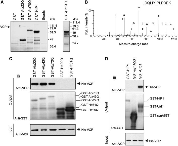
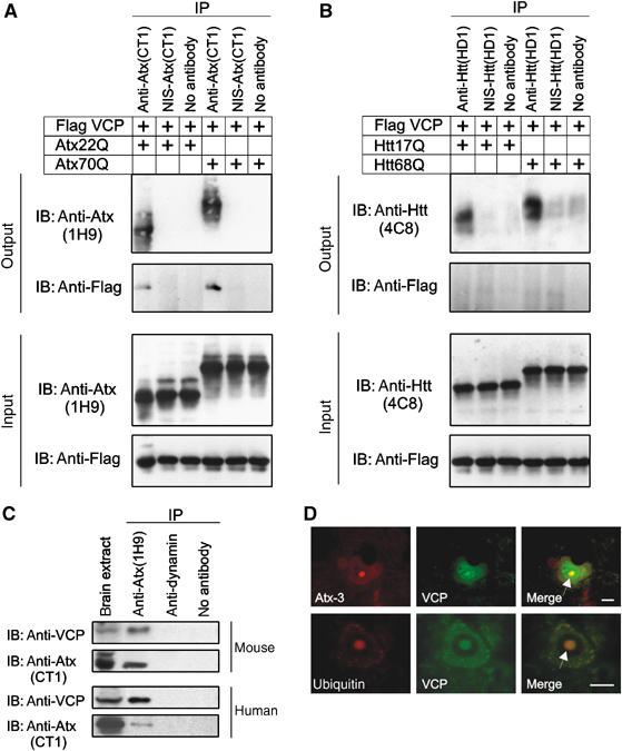
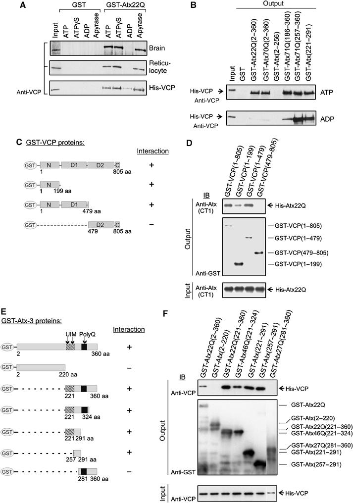
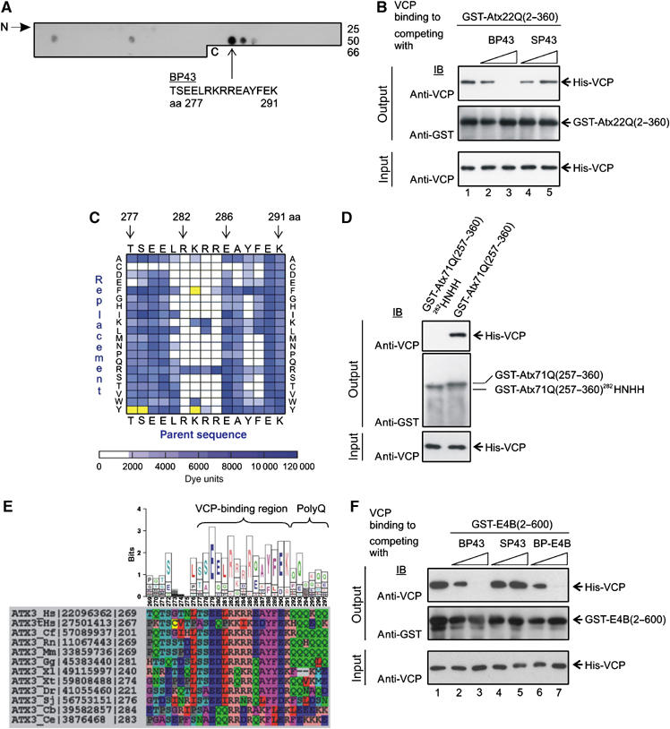
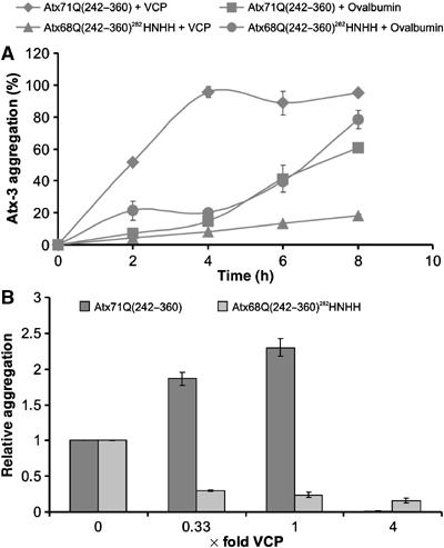
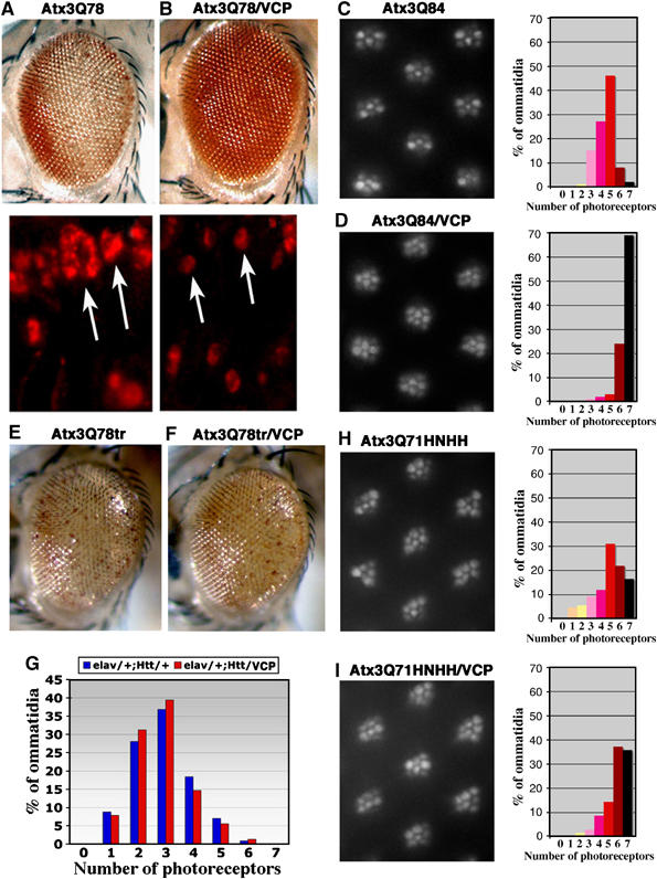
Arginine/lysine-rich motifs typically function as targeting signals for the translocation of proteins to the nucleus. Here, we demonstrate that such a motif consisting of four basic amino acids in the polyglutamine protein ataxin-3 (Atx-3) serves as a recognition site for the interaction with the molecular chaperone VCP. Through this interaction, VCP modulates the fibrillogenesis of pathogenic forms of Atx-3 in a concentration-dependent manner, with low concentrations of VCP stimulating fibrillogenesis and excess concentrations suppressing it. No such effect was observed with a mutant Atx-3 variant, which does not contain a functional VCP interaction motif. Strikingly, a stretch of four basic amino acids in the ubiquitin chain assembly factor E4B was also discovered to be critical for VCP binding, indicating that arginine/lysine-rich motifs might be generally utilized by VCP for the targeting of proteins. In vivo studies with Drosophila models confirmed that VCP selectively modulates aggregation and neurotoxicity induced by pathogenic Atx-3. Together, these results define the VCP-Atx-3 association as a potential target for therapeutic intervention and suggest that it might influence the progression of spinocerebellar ataxia type 3.


Figures

Figure 1
Figure 1
Identification and characterization of the VCP–Atx-3 protein interaction. (A) GST pulldown experiments with human brain extracts. GST fusion proteins used as baits are indicated on top of the gels. Bound proteins eluted from the affinity matrix were analysed by SDS–PAGE and Coomassie blue staining. An asterisk marks the band which was examined by mass spectrometry. (B) Fragmentation spectrum of a VCP peptide. Circles mark matches between peaks in the fragmentation spectrum and theoretical peptide masses for the identified VCP peptide. (C, D) GST pulldown experiments with purified His-VCP. Bound proteins were analyzed with an anti-VCP antibody (top panel) and bait proteins with an anti-GST antibody (middle panel). In all, 10% of the input binding mixture was also subjected to immunoblot analysis with anti-VCP antibody (bottom panel).
Figure 2
Figure 2
Analysis of the interaction between VCP and Atx-3. (A–C) Co-IP experiments with COS-1, mouse, and human brain cell extracts. Antisera used for IP and IB are indicated. The bottom panels in (A) and (B) show 10 μg of the input lysates. In (C), 50 μg of brain extracts were loaded as a control. NIS, nonimmune serum. (D) Co-immunofluorescence microscopy of SCA3 pons sections with anti-VCP (green), anti-Atx-3 (1H9, red) or anti-ubiquitin (red) antibodies. Neuronal intranuclear inclusions are indicated with an arrow. Scale bar: 10 μm (For color see web version).
Figure 3
Figure 3
Analysis of the nucleotide dependence of the Atx-3–VCP interaction and mapping of the protein-binding sites. (A, B) GST fusion proteins were incubated with lysates containing endogenous VCP or purified recombinant His-VCP in the presence or absence of nucleotides (apyrase treatment), and protein complexes were enriched with affinity beads. After extensive washing of the beads, bound protein was detected by IB using an anti-VCP antibody. Input material: 10% of brain or reticulocyte lysates or 2% His-VCP. (C, E) Schematic representation of GST-VCP and GST–Atx-3 fusion proteins and summary of binding results. (D, F) GST pulldown experiments. Binding of recombinant His-Atx22Q and His-VCP was tested against various GST-VCP and GST–Atx-3 fusion proteins, respectively. Bound proteins (top panels) as well as immobilized bait proteins (middle panels) were detected by IB using specific antibodies. The bottom panels show 10% of the input mixtures.
Figure 4
Figure 4
Identification of the amino-acid residues in Atx-3 responsible for VCP binding. (A) Detection of VCP-interacting Atx-3 peptides using overlay assays. An array with 66 overlapping 15mer peptides covering the C-terminus of Atx-3 (aa 151–360) was incubated with His-VCP, and binding peptides were identified by IB using an anti-VCP antibody. The Atx-3 binding peptide 43 (BP43) interacted most strongly with His-VCP. (B) BP43 at a concentration of 1 mM prevents the interaction between GST-Atx22Q(2–360) and His-VCP (lane 3). No competition of the protein–protein interaction was observed with the control peptide SP43. (C) Identification of the amino acids in BP43 required for the interaction with His-VCP. An array with 300 synthetic peptide analogs of BP43 with single amino-acid substitutions was incubated with His-VCP, and the amino acids critical for the peptide–protein interaction were determined by overlay assays. Signals were quantified and are represented as a spectral diagram. The BP43 parent sequence was displayed on the top and bottom of the matrix. The replaced amino acids are shown on the right and left sides. The bar below the matrix gives the signal intensity scale in arbitary dye units. As a control, anti-VCP antibody alone was incubated with the peptide array. BP43 derivatives, which were detected as false positives by the antibody alone, are indicated as yellow squares. (D) GST-pulldown experiments. The arginine/lysine-rich motif (282RKRR) in Atx-3 is critical for the interaction with His-VCP in vitro. Mutation of this sequence (282HNHH) prevents His-VCP binding. The bottom panel shows 10% of input mixture. (E) Conservation of the VCP-binding region in homologous Atx-3 sequences. The sequence logo (top) is derived from the sequence alignments of Atx-3 orthologs (bottom). Physicochemically similar amino acids are colored identical in the alignment. NCBI sequence accession numbers and the starting positions of the amino-acid sequences are indicated. Hs, Homo sapiens; tHs, Atx-3 paralog expressed in testis; Cf, Canis familiaris; Rn, Rattus norvegicus; Mm, Mus musculus; Gg, Gallus gallus; Xl/Xt, Xenopus laevis/tropicalis; Dr, Danio rerio; Sj, Schistosoma japonicum; Cb/Ce, Caenorhabditis briggsae/elegans. (F) The peptides BP43 and BP-E4B at a concentration of 1 mM prevent the interaction between GST-E4B(2–600) and His-VCP (lanes 3 and 7). The control peptide SP43 has no effect.
Figure 5
Figure 5
Binding of VCP to Atx-3 influences the assembly of polyQ-containing protein aggregates. (A) VCP stimulates the formation of Atx-3 aggregates in vitro. 3 μM GST-Atx71Q(242–360) or GST-Atx68Q(242–360)282HNHH was incubated with 1 μM VCP or 1 μM ovalbumin in the presence of PP. The assembly of SDS-stable Atx-3 protein aggregates was monitored by filtration using an anti-Atx (CT1) antibody. The sample with the highest signal intensity was arbitrarily set as 100%. (B) Effect of different VCP concentrations on Atx-3 aggregation in vitro. GST fusion proteins were incubated for 6 h with PP and different amounts of VCP. The assembly of SDS-insoluble Atx71Q(242–360) and Atx68Q(242–360)282HNHH aggregates was monitored by filtration using the CT1 antibody. Relative amounts of aggregates were quantified by phosphorimager densitometry. The X-axis of the diagram shows the ratio of VCP to Atx-3.
Figure 6
Figure 6
Human VCP suppresses Atx-3-induced neurodegeneration in Drosophila. (A) Flies expressing pathogenic Atx3Q78 protein alone (top is the eye, bottom are protein accumulations in adult retinal cryosections) or (B) together with VCP. Degeneration of the eye and size of protein aggregates are mitigated with added VCP activity. Arrows highlight nuclear inclusions. (C) Photographs of ommatidia from 2-day-old flies expressing Atx3Q84 alone or (D) with VCP, with distribution (in percentage) of photoreceptor neurons in ommatidia, with 7 being normal. Mean number of photoreceptor neurons per ommatidium is 4.5 in (C) and 6.6 in (D), indicating an improvement of 30% with added VCP. Genotypes: (A) gmr-Gal4;UAS-Atx3Q78#24.1/+, (B) gmr-Gal4;UAS-Atx3Q78#24.1/+;UAS-VCP/+, (C) elav-Gal4/+;UAS-Atx3Q84#16.1/+, (D) elav-Gal4/+;UAS-Atx3Q84#16.1/+;UAS-VCP/+. The VCP/Atx-3 ratio is ∼1.45 for Atx-3 line #24.1 and ∼4.2 for line #16.1. (E, F) Coexpression of VCP has little effect on degeneration induced by a truncated version of Atx-3 with a shortened VBM domain. Flies expressing Atx3Q78tr (E) alone or (F) with VCP. Genotypes: gmr-GAL4;UAS-Atx3Q78tr(S)/+ (E) or in trans to UAS-VCP (F). (G) Coexpression of VCP has little effect on degeneration induced by pathogenic Htt protein. Distribution in percentage of photoreceptor neurons in ommatidia of flies expressing HttQ120 alone or with VCP. Genotypes: elav-Gal4/+;gmr-HttQ120/+ (blue) and elav-Gal4/+;gmr-HttQ120/UAS-VCP (red). Photographs of ommatidia from 14-day-old flies expressing Atx3Q71HNHH (H) alone or with VCP (I), with distribution (in percentage) of photoreceptor neurons in ommatidia, with 7 being normal. Mean number of photoreceptors per ommatidium is 4.9 in (H) and 5.9 in (I), indicating an improvement of only 14% by VCP. Thus, photoreceptor neuron degeneration in flies expressing Atx3Q71HNHH is partially mitigated by VCP, but only to half the extent as degeneration by pathogenic Atx-3 with an intact VBM.

Similar articles

Cited by

References

    1. Albrecht M, Golatta M, Wullner U, Lengauer T (2004) Structural and functional analysis of ataxin-2 and ataxin-3. Eur J Biochem 271: 3155–3170 - PubMed
    1. Bevivino AE, Loll PJ (2001) An expanded glutamine repeat destabilizes native ataxin-3 structure and mediates formation of parallel beta-fibrils. Proc Natl Acad Sci USA 98: 11955–11960 - PMC - PubMed
    1. Burnett B, Li F, Pittman RN (2003) The polyglutamine neurodegenerative protein ataxin-3 binds polyubiquitylated proteins and has ubiquitin protease activity. Hum Mol Genet 12: 3195–3205 - PubMed
    1. Chai Y, Koppenhafer SL, Shoesmith SJ, Perez MK, Paulson HL (1999) Evidence for proteasome involvement in polyglutamine disease: localization to nuclear inclusions in SCA3/MJD and suppression of polyglutamine aggregation in vitro. Hum Mol Genet 8: 673–682 - PubMed
    1. Chernoff YO, Lindquist SL, Ono B, Inge-Vechtomov SG, Liebman SW (1995) Role of the chaperone protein Hsp104 in propagation of the yeast prion-like factor [psi+]. Science 268: 880–884 - PubMed

Publication types

MeSH terms