. 2006 Mar 22;25(6):1177-83.
doi: 10.1038/sj.emboj.7601028. Epub 2006 Mar 9.

Structural evidence for induced fit and a mechanism for sugar/H+ symport in LacY

Affiliations

Structural evidence for induced fit and a mechanism for sugar/H+ symport in LacY

Osman Mirza et al. EMBO J. .

Erratum in

  • EMBO J. 2006 May 3;25(9):2038

Abstract

Cation-coupled active transport is an essential cellular process found ubiquitously in all living organisms. Here, we present two novel ligand-free X-ray structures of the lactose permease (LacY) of Escherichia coli determined at acidic and neutral pH, and propose a model for the mechanism of coupling between lactose and H+ translocation. No sugar-binding site is observed in the absence of ligand, and deprotonation of the key residue Glu269 is associated with ligand binding. Thus, substrate induces formation of the sugar-binding site, as well as the initial step in H+ transduction.


Figures

Figure 1
Figure 1
Overall structure of C154G LacY in the absence of ligand. LacY consists of 12 transmembrane helices organized in two pseudo-symmetrical α-helical bundles. The N- and C-terminal 6-helix domains form an internal cavity open to the cytoplasmic side. In the ligand-free structure at pH 6.5, the residues involved in substrate binding (Glu126 (helix IV), Arg144 and Trp151 (helix V)) are shown in yellow, and those involved in H+ translocation [Tyr236 (helix VII), Arg302 (helix IX), His322 and Glu325 (helix X)] are shown in pink. The dual-function residue Glu269 (helix VIII) is shown in cyan. (A) Side view; (B) cytoplasmic view.
Figure 2
Figure 2
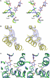
Re-arrangement of the active site in the absence and presence of sugar. (A) Cytoplasmic stereo view of the sugar-binding site from superimposed crystal structures. The TDG-bound structure (1pv7) is shown in blue (3); ligand-free structures are shown in green (pH 5.6) and in yellow (pH 6.5). (B) Residues involved in sugar binding are shown with a 2Fo−Fc sigmaA weighted electron density map from the structure obtained at pH 6.5. (C) Stereo view of Glu269 in a relatively hydrophobic environment surrounded by residues Trp151, Cys148, Asn272, Ala273 and Met323.
Figure 3
Figure 3
Stereo view of residues involved in H+ translocation network. (A) Residues involved in H+ translocation in the TDG-bound structure (blue; 1pv7) and ligand-free structure (green) at pH 5.6. (B) Side-chains involved in H+ translocation at pH 5.6 in the absence of ligand shown with a 2Fo−Fc sigmaA weighted electron density map.
Figure 4
Figure 4
Proposed model for the initial step in coupling between sugar and H+ translocation. H-bonds are represented by broken lines; charge pairs by solid lines. The proton is represented by a white sphere and the water by a turquoise sphere. (A) Glu269 is protonated and close to Trp151 to form a platform in the absence of substrate. Arg144 forms a salt bridge with Glu269. (B) The Glu269/Trp151 platform orients the galactopyranosyl ring to allow specific interactions with Arg144, Glu126 and Glu269. (C) Arg144 moves towards the galactopyranoside ring to form a bi-dentate H-bond with the O4 and O3 positions. Glu269 moves into a hydrophilic environment, deprotonates and interacts with the O3 position of the sugar, as well as a salt bridge with Arg144. (D) The H+ is transferred to His322 via water.
Figure 5
Figure 5
An updated influx mechanism for lactose/H+ symport. See main text for details. The proton (H+) and the substrate (S) are shown with red and green, respectively. Charge pairs are indicated by solid lines.

References

    1. Abramson J, Smirnova I, Kasho V, Verner G, Kaback HR, Iwata S (2003) Structure and mechanism of the lactose permease of Escherichia coli. Science 301: 610–615 - PubMed
    1. Brünger A, Adams P, Clore G, DeLano W, Gros P, Grosse-Kunstleve R, Jiang J, Kuszewski J, Nilges M, Pannu N, Read R, Rice L, Simonson T, Warren G (1998) Crystallography & NMR Systems: a new software suite for macromolecular structure determination. Act Crystallogr D54: 905–921 - PubMed
    1. Frillingos S, Kaback HR (1997) The role of helix VIII in the lactose permease of Escherichia coli. II. Site-directed sulfhydryl modification. Protein Sci 6: 438–443 - PMC - PubMed
    1. Frillingos S, Sahin-Toth M, Wu J, Kaback HR (1998) Cys-scanning mutagenesis: a novel approach to structure function relationships in polytopic membrane proteins. FASEB J 12: 1281–1299 - PubMed
    1. Guan L, Hu Y, Kaback HR (2003) Aromatic stacking in the sugar binding site of the lactose permease. Biochemistry 42: 1377–1382 - PubMed

Publication types

LinkOut - more resources