. 2006 Apr;87(Pt 4):855-860.
doi: 10.1099/vir.0.81664-0.

Core protein cleavage by signal peptide peptidase is required for hepatitis C virus-like particle assembly

Affiliations

Core protein cleavage by signal peptide peptidase is required for hepatitis C virus-like particle assembly

Malika Ait-Goughoulte et al. J Gen Virol. 2006 Apr.

Abstract

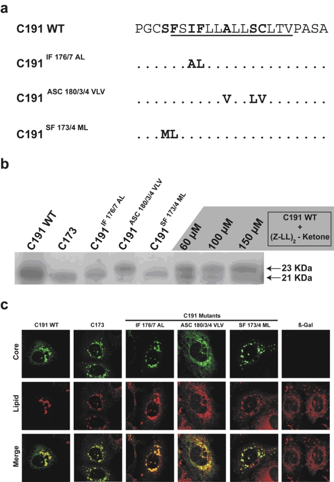
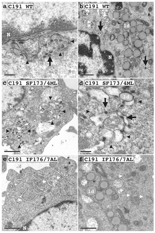
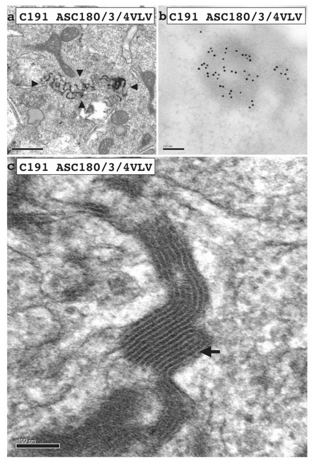
Hepatitis C virus (HCV) core protein, expressed with a Semliki Forest virus replicon, self-assembles into HCV-like particles (HCV-LP) at the endoplasmic reticulum (ER) membrane, providing an opportunity to study HCV assembly and morphogenesis by electron microscopy. This model was used to investigate whether the processing of the HCV core protein by the signal peptide peptidase (SPP) is required for the HCV-LP assembly. Several mutants were designed as there are conflicting reports concerning the cleavage of mutant proteins by SPP. Production of the only core mutant protein that escaped SPP processing led to the formation of multiple layers of electron-dense ER membrane, with no evidence of HCV-LP assembly. These data shed light on the HCV core residues involved in SPP cleavage and suggest that this cleavage is essential for HCV assembly.


Figures

Fig. 1
Fig. 1
Analysis of HCV core-E1 signal peptide processing, (a) Signal sequences of WT C191 HCV core protein and mutants (IF176/7AL, ASC180/3/4VLV and SF173/4ML). The transmembrane region of the signal sequence is underlined, (b): Production in BHK-21 cells of the WT and mutant C191 proteins and analysis of their processing by SDS-PAGE and western blotting, using the human monoclonal anti-HCV core antibody B12F8 (Esposito et al., 1995). The mutants were compared with the WT C191 protein in the absence or presence of various concentrations of the SPP inhibitor (Z-LL)2-ketone, and with the reference core protein C173. (c). Immunofluorescence of WT and mutant HCV core proteins combined with Nile Red staining of lipid droplets in FLC4 cells.
Fig. 2
Fig. 2
Electron micrographs of BHK-21 cells electroporated with WT C191 (a & b), C191 SF173/4ML (c & d), or C191 IF176/7AL (e & f) RNA. Micrograph in a shows a zone of convoluted ER membranes (delimited by arrowheads) in which HCV-LP assembly (arrows) was frequent. Micrograph in b shows that clusters of large lipid droplets were found in the perinuclear area next to ER membranes in which HCV-LP assembly (large black arrows) was detected. Cells transfected with C191 SF173/4ML RNA presented a pattern similar to that of the WT protein, with convoluted ER membranes (delimited by arrowheads in c) in which HCV-LP assembly (arrows in d) was detected. Cells transfected with C191 IF176/7AL RNA had normal ER structures, evenly distributed throughout the cytoplasm (white arrows), in which no HCV-LP was detected, as for cells transfected with β-Gal recombinant RNA (not shown). The only subtle change in ultrastructure of cells producing C191 IF176/7AL was the clustering of lipid droplets in the perinuclear area, next to normal ER membranes (micrograph in f). N, nucleus. ld: lipid droplet. ER lu: ER lumen. Bars in a and d, 0,2 μm. Bars in b, c, e and f, 1 μm.
Fig. 3
Fig. 3
Electron micrographs of BHK-21 cells electroporated with the C191 ASC180/3/4VLV RNA, showing electron-dense ER membranes (delimited by arrowheads at low magnification in a). At high magnification (c), these structures were found to be formed by the interaction of multiple ER membranes (arrow). In b, an immunogold labeling with anti-core antibodies demonstrated the presence of high amount of core protein in these multi-layered ER membranes. Bαρσιν α, 0,2 μm. Bars in b and c, 100 nm.

Similar articles

Cited by

References

    1. Aizaki H, Nagamori S, Matsuda M, et al. Production and release of infectious hepatitis C virus from human liver cell cultures in the three-dimensional radial-flow bioreactor. Virology. 2003;314:16–25. - PubMed
    1. Blanchard E, Brand D, Trassard S, Goudeau A, Roingeard P. Hepatitis C virus-like particle morphogenesis. J Virol. 2002;76:4073–4079. - PMC - PubMed
    1. Blanchard E, Hourioux C, Brand D, Ait-Goughoulte M, Moreau A, Trassard S, Sizaret PY, Dubois F, Roingeard P. Hepatitis C virus-like particle budding: role of the core protein and importance of its Asp 111. J virol. 2003;77:10131–10138. - PMC - PubMed
    1. Edbauer D, Winkler E, Regula JT, Pesold B, Steiner H, Haass C. Reconstitution of gamma-secretase activity. Nat Cell Biol. 2003;5:486–488. - PubMed
    1. Esposito G, Scarselli E, Cerino A, Mondelli MU, La Monica N, Traboni C. A human antibody specific for hepatitis C virus core protein: synthesis in a bacterial system and characterization. Gene. 1995;164:203–209. - PubMed

Publication types

Substances

LinkOut - more resources