. 2006 Apr;116(4):878-91.
doi: 10.1172/JCI26323. Epub 2006 Mar 9.

Disease-associated mutations affect intracellular traffic and paracellular Mg2+ transport function of Claudin-16

Affiliations

Disease-associated mutations affect intracellular traffic and paracellular Mg2+ transport function of Claudin-16

P Jaya Kausalya et al. J Clin Invest. 2006 Apr.

Abstract

Claudin-16 (Cldn16) is selectively expressed at tight junctions (TJs) of renal epithelial cells of the thick ascending limb of Henle's loop, where it plays a central role in the reabsorption of divalent cations. Over 20 different mutations in the CLDN16 gene have been identified in patients with familial hypomagnesemia with hypercalciuria and nephrocalcinosis (FHHNC), a disease of excessive renal Mg2+ and Ca2+ excretion. Here we show that disease-causing mutations can lead to the intracellular retention of Cldn16 or affect its capacity to facilitate paracellular Mg2+ transport. Nine of the 21 Cldn16 mutants we characterized were retained in the endoplasmic reticulum, where they underwent proteasomal degradation. Three mutants accumulated in the Golgi complex. Two mutants were efficiently delivered to lysosomes, one via clathrin-mediated endocytosis following transport to the cell surface and the other without appearing on the plasma membrane. The remaining 7 mutants localized to TJs, and 4 were found to be defective in paracellular Mg2+ transport. We demonstrate that pharmacological chaperones rescued surface expression of several retained Cldn16 mutants. We conclude that FHHNC can result from mutations in Cldn16 that affect intracellular trafficking or paracellular Mg2+ permeability. Knowledge of the molecular defects associated with disease-causing Cldn16 mutations may open new venues for therapeutic intervention.


Figures

Figure 1
Figure 1. Predicted topology of Cldn16 and location of the different mutations linked to FHHNC reported.
Shown is the amino acid sequence in 1-letter code, with mutated residues in yellow and arrows indicating the change introduced by the mutation. The effect of the corresponding mutation on the predominant steady-state distribution of Cldn16 is highlighted in different colors: green, cell surface; red, ER; dark blue, Golgi complex; light blue, lysosomes. X, stop codon, fs, frame shift. The peptide (T52-S66) used to generate the anti-loop antibody is shaded in gray.
Figure 2
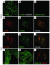
Figure 2. Clathrin-mediated endocytosis of Cldn16.
(AC) Characterization of an antibody to the first extracellular loop of Cldn16 (α-loop) and endogenous Cldn16 expression in MDCK cells. MDCK cells were incubated for 1 hour at 37°C in the presence of anti-loop antibody alone (A), together with antigenic peptide (B), or with preimmune serum (C). Cells were then washed, fixed, and permeabilized, and the anti-loop antibody was detected by immunofluorescence microscopy using a labeled secondary antibody. (DF) Detection of HA-tagged Cldn16 expressed in MDCK cells. MDCK cells expressing N-terminally HA epitope–tagged Cldn16 were incubated for 1 hour at 37°C with anti-loop antibody. α-HA (D) and the anti-loop antibody (E) were then detected in permeabilized cells. (F) Merging the HA and anti-loop antibody staining shows extensive colocalization at the cell surface and in a few intracellular vesicles. (GI) Detection of anti-loop antibody internalized via HA-tagged Cldn16 in early endosomes of transfected HeLa cells. Anti-loop antibody (G) and EEA1 (H) were visualized by immunofluorescence microscopy, and the 2 images were merged (I). (JL) Clathrin-mediated internalization of Cldn16. HeLa cells expressing HA-tagged Cldn16 were incubated for 1 hour at 37°C with anti-loop antibody under either hypertonic (J) or cytosol acidified (K) conditions to disrupt clathrin function or in the presence of cholesterol oxidase (L) to disrupt caveolae. Cldn16 was then visualized by immunofluorescence microscopy. In L, cells were stained with antibodies to EEA1, and the merged image is shown. Shown are representative images of 2–3 independent experiments.
Figure 3
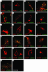
Figure 3. Cell surface expression of Cldn16 mutants linked to FHHNC.
HeLa cells transiently expressing the indicated HA-tagged Cldn16 mutants were incubated in the presence of anti-loop antibody for 1 hour at 37°C and then immunostained to detect the HA tag (red) or the anti-loop antibody (green). Merged images are shown. Shown are representative images of 2–3 independent experiments.
Figure 4
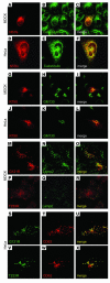
Figure 4. Steady-state localization of selected Cldn16 mutants to different subcellular organelles.
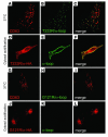
(AF) Example of a representative mutant with predominant ER localization. MDCK or HeLa cells expressing HA-tagged N53fs were immunostained for HA (A and D, red) and the ER marker calreticulin (B and E, green). Colocalization was apparent in the merged images (C and F, yellow). (GL) Representative mutant with predominant Golgi localization. MDCK or HeLa cells expressing HA-tagged R79X were immunostained for HA (G and J, red) and the Golgi marker GM130 (H and K, green). Colocalization was apparent in the merged images (I and L, yellow). (MX) Mutants with predominant lysosomal localization. MDCK or HeLa cells expressing HA-tagged G121R or T233R were immunostained for HA (M and P, red, and S and V, green) and the lysosome markers Lamp2 (N and Q, green) or CD63 (T and W, red). Colocalization was apparent in the merged images (O, R, U, and X, yellow). Shown are representative images of 2–3 independent experiments. For the remaining mutants, see Supplemental Figure 1.
Figure 5
Figure 5. Characterization of intracellular trafficking defects of representative Cldn16 mutants.
Transfected MDCK cells expressing the indicated HA-tagged Cldn16 mutants were incubated at 20°C for 3 hours to allow transport out of the ER but prevent exit from the Golgi complex and TGN. Cells were then either processed for immunofluorescence staining (A and D) or transferred to 37°C for 1 hour (B and E) or 6 hours (C and F) in the presence of cycloheximide and ALLN to inhibit de novo protein synthesis and degradation, respectively. The cells were then immunostained with antibodies to HA to detect the tagged Cldn16 mutant (red) and either the Golgi marker GM130 (A, B, D, and E, green) or the lysosomal membrane protein CD63 (C and F, green). Shown are representative images of 2–3 independent experiments. For the remaining mutants, see Supplemental Figure 2.
Figure 6
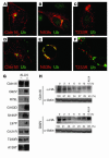
Figure 6. ER-retained Cldn16 mutants are subject to proteasomal degradation.
(AF) Colocalization of ER-retained Cldn16 mutants with ubiquitin was increased in the presence of a proteasome inhibitor. Transfected MDCK cells expressing HA-tagged Cldn16 or the indicated mutants were incubated for 10 hours in the absence (AC) or presence (DF) of the proteasome inhibitor ALLN. Cells were then immunostained with antibodies to detect the HA-tagged Cldn16 mutant (red) or ubiquitin (Ub, green). Colocalization was apparent in the merged images (yellow). Shown are representative images of 2–3 independent experiments. For the remaining mutants, see Supplemental Figure 3. (G) ER-retained Cldn16 mutants were stabilized by proteasome inhibitors. HEK-293T cells transiently expressing the indicated HA-tagged Cldn16 mutants were incubated in the absence (–) or presence (+) of the proteasome inhibitor ALLN. Cells were then lysed, and Cldn16 was detected by SDS-PAGE and Western blot analysis. Blotting for actin was used as a loading control (data not shown). (H) Turnover of ER-retained Cldn16 mutants was blocked by proteasome inhibitors. HEK-293T cells transiently expressing HA-tagged Cldn16 or the ER-retained HA-tagged G92V were cultured in cycloheximide to block de novo protein synthesis for the time periods indicated, either in the absence or presence of the proteasome inhibitor ALLN. Cells were then lysed, and Cldn16 detected by SDS-PAGE and Western blot analysis. Percent expression relative to that at 0 hours is shown below each lane. Blotting for actin was used as a loading control. One representative blot and quantification of 3 independent experiments is shown.
Figure 7
Figure 7. Cldn16 mutants that localize to lysosomes follow different pathways.
Transfected HeLa cells expressing HA-tagged T233R (AF) or G121R (GL) were incubated in the presence of anti-loop antibodies at 37°C for 1 hour, either under normal conditions (AC and GI) or cytosol acidification to block endocytosis (DF and JL). The cells were then immunostained with antibodies to detect the anti-loop antibody (B, E, H, and K; green), the HA-tagged Cldn16 mutant (D and J; red), or the lysosomal membrane protein CD63 (A and G; red). Colocalization was apparent in the merged images (C, F, I, and L; yellow). Shown are representative images of 2–3 independent experiments.
Figure 8
Figure 8. Chemical chaperones rescue cell surface expression of several Cldn16 mutants.
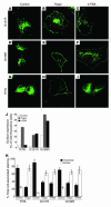
Transfected HeLa cells expressing HA-tagged G121R (AC), G169R (DF), or R79L (GI) were incubated in the presence of vehicle (Control; A, D, and G), thapsigargin (Thaps; B, E, and H) or 4-PBA (C, F, and I). The cells were then immunostained with antibodies to detect the Cldn16 mutant (green). Shown are representative images of 2–3 independent experiments. (J) Quantification of the number of cells that expressed mutant Cldn16 on the cell surface in response to thapsigargin or 4-PBA. One hundred randomly chosen cells expressing the Cldn16 mutant were analyzed, and the fraction of cells showing surface expression in the presence of the pharmacological chaperones was determined. Data from 3 independent experiments is shown. (K) Quantification of the fraction of total cell-associated mutant Cldn16 present on the surface of individual cells in response to thapsigargin or 4-PBA. Integrated fluorescence densities of either the entire or only the intracellular cell surface area of 50–60 individual, randomly selected cells labeled with anti-HA antibodies were determined. The density associated with the cell surface was estimated from the difference of the 2 values and normalized to total cell-associated staining. Given that only the fluorescence associated with the outline of the cells was defined as surface staining, these results likely underestimate the effect of the molecular chaperones.
Figure 9
Figure 9. TJ localization of Cldn16 mutants expressed on the cell surface.
Transfected MDCK cells expressing Cldn16 mutants that are delivered to the cell surface were grown on permeable polycarbonate filters to obtain polarized cell monolayers. The cells were then fixed, permeabilized, and stained with antibodies to HA to detect the Cldn16 mutants (green) or the TJ marker ZO-1 (red). Shown are representative images of 2–3 independent experiments.
Figure 10
Figure 10. Measurements of PMg2+ , Rt , and PNa /PCl ratios.
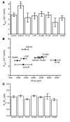
(A) PMg2+. Monolayers of stable transfected MDCK-C7 cells were grown on permeable supports and mounted in Ussing chambers. Mg2+ was added to the basolateral side, and net flux was measured by means of AAS. *P < 0.05 versus control. (B) Comparison of Rt to PMg2+. Rt of the clones ranged between 941 ± 174 Ω cm2 (T233R) and 3,406 ± 303 Ω cm2 (H71D). No correlation of PMg2+ and Rt was observed (n = 7–16). (C) PNa/PCl ratios. Dilution potentials were measured with modified Ringer’s solution on the luminal or serosal side, and the data from the 2 conditions was pooled (n = 6 per group). No significant changes were observed in cells expressing WT Cldn16 or the mutants indiced.

References

    1. Asplin J.R., Favus M.J., Coe F.L. In The kidney. 5th edition. B.M. Brenner, editor. W.B. Saunders.; Philadelphia, Pennsylvania, USA.: 1996. Nephrolithiasis. pp. 1893–1935.
    1. Weber S., et al. Familial hypomagnesemia with hypercalciuria and nephrocalcinosis maps to chromosome 3q27 and is associated with mutations in the PCLN-1 gene. Eur. J. Hum. Genet. 2000;8:414–422. - PubMed
    1. Blanchard A., et al. Paracellin-1 is critical for magnesium and calcium reabsorption in the human thick ascending limb of Henle. Kidney Int. 2001;59:2206–2215. - PubMed
    1. Simon D.B., et al. Paracellin-1, a renal tight junction protein required for paracellular Mg2+ resorption. Science. 1999;285:103–106. - PubMed
    1. Weber S., et al. Primary gene structure and expression studies of rodent paracellin-1. J. Am. Soc. Nephrol. 2001;12:2664–2672. - PubMed

Publication types

MeSH terms