. 2006 Mar 10:7:10.
doi: 10.1186/1471-2199-7-10.

Sp1/3 and NF-1 mediate basal transcription of the human P2X1 gene in megakaryoblastic MEG-01 cells

Affiliations

Sp1/3 and NF-1 mediate basal transcription of the human P2X1 gene in megakaryoblastic MEG-01 cells

Jiangqin Zhao et al. BMC Mol Biol. .

Abstract

Background: P2X1 receptors play an important role in platelet function as they can induce shape change, granule centralization and are also involved in thrombus formation. As platelets have no nuclei, the level of P2X1 expression depends on transcriptional regulation in megakaryocytes, the platelet precursor cell. Since nothing is known about the molecular mechanisms regulating megakaryocytic P2X1 expression, this study aimed to identify and functionally characterize the P2X1 core promoter utilized in the human megakaryoblastic cell line MEG-01.

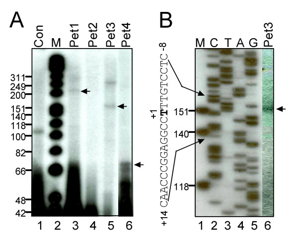
Results: In order to identify cis-acting elements involved in the transcriptional regulation of P2X1 expression, the ability of 4.7 kb P2X1 upstream sequence to drive luciferase reporter gene expression was tested. Low promoter activity was detected in proliferating MEG-01 cells. This activity increased 20-fold after phorbol-12-myristate-13-acetate (PMA) induced differentiation. A transcription start site was detected 365 bp upstream of the start codon by primer extension. Deletion analysis of reporter constructs indicated a core promoter located within the region -68 to +149 bp that contained two Sp1 sites (named Sp1a and Sp1b) and an NF-1 site. Individual mutations of Sp1b or NF-1 binding sites severely reduced promoter activity whereas triple mutation of Sp1a, Sp1b and NF-1 sites completely abolished promoter activity in both untreated and PMA treated cells. Sp1/3 and NF-1 proteins were shown to bind their respective sites by EMSA and interaction of Sp1/3, NF-1 and TFIIB with the endogenous P2X1 core promoter in MEG-01 cells was demonstrated by chromatin immunoprecipitation. Alignment of P2X1 genes from human, chimp, rat, mouse and dog revealed consensus Sp1a, Sp1b and NF-1 binding sites in equivalent positions thereby demonstrating evolutionary conservation of these functionally important sites.

Conclusion: This study has identified and characterized the P2X1 promoter utilized in MEG-01 cells and shown that binding of Sp1/3 and NF-1 to elements in the direct vicinity of the transcription start site is essential for basal transcription. Targeting the function of these transcription factors in megakaryocytes may therefore provide a basis for the future therapeutic manipulation of platelet P2X1 function.


Figures

Figure 1
Figure 1
The P2X1 transcription start site is located 365 bp upstream of the start codon. A) 32P-labelled primers Pet1, Pet2, Pet3 and Pet4 and Con (control primer pTXRHR-100) were annealed to poly(A) RNA from PMA-treated MEG-01 cells and extended with reverse transcriptase. Extension products for Pet1, Pet3 and Pet4 that equate to the same transcription start site are indicated with arrows. (M), molecular mass marker. B) Pet3 extension reaction (lane 6) run in parallel with a sequencing ladder. The location of primers is depicted in Figure 3.
Figure 2
Figure 2
Activity of P2X1 promoter constructs in transient transfection assays. A) Promoter activity of p-4407/+365 was determined both with (+) and without (-) the prior treatment of cells with 10 nM PMA. Data are means of absolute firefly luciferase activity of three independent transfections. B) Luciferase activity of P2X1 deletion constructs in PMA treated MEG-01 cells. TSS, transcription start site. PT depicts a polypyrimidine tract located from -1801 to -1405 bp. Values are presented as means of at least three experiments and normalized against the construct p-4407/+365. Names of plasmid constructs correspond to the 5' and 3' ends of the insert relative to the transcription start site e.g. construct p-4407/+365 extends from 4407 bp 5' to 365 bp 3' of the transcription start site.
Figure 3
Figure 3
Sequence of the P2X1 proximal promoter region. Potential transcription factor binding sites are underlined and in bold. Oligonucleotide sequences used for primer extension analysis (Pet1-4) are indicated by horizontal arrows. End points of plasmid deletion constructs in this region are indicated by "p" and number above the sequence. Sequence numbering is relative to the transcription start site indicated as +1. The translation initiation ATG is boxed. The EMBL/Genbank accession number of this sequence is AJ971536.
Figure 4
Figure 4
EMSA assays on NF-1 and Sp1/Sp3 binding sites. A) Competition experiments were performed in the presence of excess unlabelled O-NF1 (lane 4) and unlabelled mutated O-NF1-mut (lane 5). Complexes supershifted by pre-incubation with an NF-1 antibody (NF-1Ab, lane 2) are marked "SS". "NS" indicates non specific binding complexes, the level of which varied between individual nuclear extractions (lanes 1 and 3). "F" indicates free probe. B) Supershift assays using Sp1 (lanes 2 and 5) and Sp3 (lanes 3 and 6) specific antibodies. C) Competition experiments in the absence (lanes 1 and 5) or presence of excess of unlabelled competitors: Lanes 2 and 6, an excess of unlabelled O-Sp1a or O-Sp1b probe respectively. Lanes 3 and 7, an excess of unlabelled mutated oligonucleotide O-Sp1a-mut and O-Sp1b-mut respectively. Lanes 4 and 8, an excess of a consensus Sp1 oligonucleotide (Promega, E323A).
Figure 5
Figure 5
Mutation analysis of transcription factor binding sites. A series of luciferase reporter constructs harbouring site-directed mutations in the NF-κB, NF-1 and Sp1 binding sites of the P2X1 proximal promoter region were assayed for promoter activity in PMA-treated MEG-01 cells (sequences of mutations are shown in Table 2). Shaded symbols represent mutated binding sites. Values are presented as means ± S.E of at least three independent experiments and are normalized against the wild type p-190/+149 construct for panels A to C and the wild type construct p-4407/+365 in panel D. A) Effects of mutations in the construct p-190/+149. B) Effect of mutation after removal of the potential NF-κB binding site. C) Effect of mutation after removal of the transcription start site that is utilized by smooth muscle cells [15]. Dotted vertical arrow (TSS) represents the transcription start site determined in this study. D) The Sp1a, Sp1b and NF-1 sites were mutated in the original full length 4.7 kb reporter construct p-4407/+365 to generate the triple mutated construct p-4407/+365(mut4). Promoter activities were determined in MEG-01 cells either treated (+) or untreated (-) with PMA.
Figure 6
Figure 6
Transcription factors bind the endogenous P2X1 promoter. Formaldehyde cross-linked chromatin was prepared from PMA-treated MEG-01 cells and immunoprecipitated with antibodies to Sp1 (lane 4), Sp3 (lane 5), NF-1 (lane 6), TFIIB (lane 7), or in the absence of antibody (lane 3). PCR was performed with specific primers for the P2X1, H4 and Hsp70 promoter and for the GAPDH coding region as a negative control. A sample representative of the total input chromatin (input DNA lane 1) was included in the PCR analysis. Lane 2 shows the supernatant of the 'unbound' sample. PCR products were between 282 and 983 bp in length. PCR cycle numbers were 31 for the P2X1, H4 and Hsp70 promoter and 36 for the GAPDH coding region. Primer sequences are given in Table 3.
Figure 7
Figure 7
Cross species conservation of the P2X1 core promoter. P2X1 upstream sequence from chimp (chromosome 19 position 4051445–4051944), dog (chromosome 9 position 40295005–40295504), rat (chromosome 10 position 59889637–59890136) and mouse (chromosome 11 position 72611373–72611872) was obtained by BLAST searching of genomic databases via the ENSEMBL genome browser . The 500 bp upstream of the ATG start codon of each gene was aligned using CLUSTAL. Alignment equivalent to -68/+149 of the human P2X1 core promoter is shown. Sequence identity to the human P2X1 sequence over this region was 99.1%, 83.4%, 76.8%, and 75.9% for chimp, dog, mouse and rat respectively. "." indicates nucleotides identical to the human P2X1 sequence. Dashes indicate positions where gaps have been inserted in order to maintain the alignment. The transcription start site of the human P2X1 gene determined in this study is indicated as +1. Potential transcription start sites predicted by PROSCAN [17] are indicated as arrows. In each species, potential NF-1, Sp1a and Sp1b binding sites (indicated by boxes) are present in equivalent positions to the human P2X1 core promoter.

References

    1. North RA. Molecular physiology of P2X receptors. Physiol Rev. 2002;82:1013–1067. - PubMed
    1. Barrera NP, Ormond SJ, Henderson RM, Murrell-Lagnado RD, Edwardson JM. Atomic force microscopy imaging demonstrates that P2X2 receptors are trimers but that P2X6 receptor subunits do not oligomerize. J Biol Chem. 2005;280:10759–10765. doi: 10.1074/jbc.M412265200. - DOI - PubMed
    1. Nicke A, Baumert HG, Rettinger J, Eichele A, Lambrecht G, Mutschler E, Schmalzing G. P2X1 and P2X3 receptors form stable trimers: a novel structural motif of ligand-gated ion channels. Embo J. 1998;17:3016–3028. doi: 10.1093/emboj/17.11.3016. - DOI - PMC - PubMed
    1. Valera S, Hussy N, Evans RJ, Adami N, North RA, Surprenant A, Buell G. A new class of ligand-gated ion channel defined by P2x receptor for extracellular ATP. Nature. 1994;371:516–519. doi: 10.1038/371516a0. - DOI - PubMed
    1. Vial C, Evans RJ. P2X(1) receptor-deficient mice establish the native P2X receptor and a P2Y6-like receptor in arteries. Mol Pharmacol. 2002;62:1438–1445. doi: 10.1124/mol.62.6.1438. - DOI - PubMed

Publication types

MeSH terms