. 2006 Mar 10;124(5):997-1009.
doi: 10.1016/j.cell.2005.12.038.

Ligands for clathrin-mediated endocytosis are differentially sorted into distinct populations of early endosomes

Affiliations

Ligands for clathrin-mediated endocytosis are differentially sorted into distinct populations of early endosomes

Melike Lakadamyali et al. Cell. .

Abstract

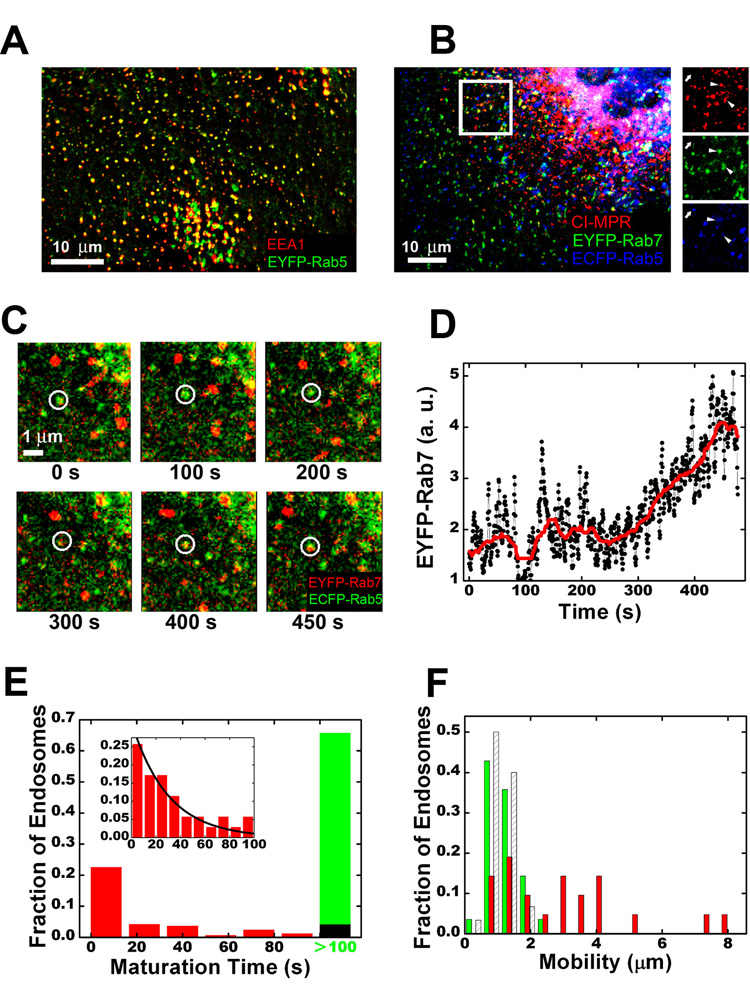
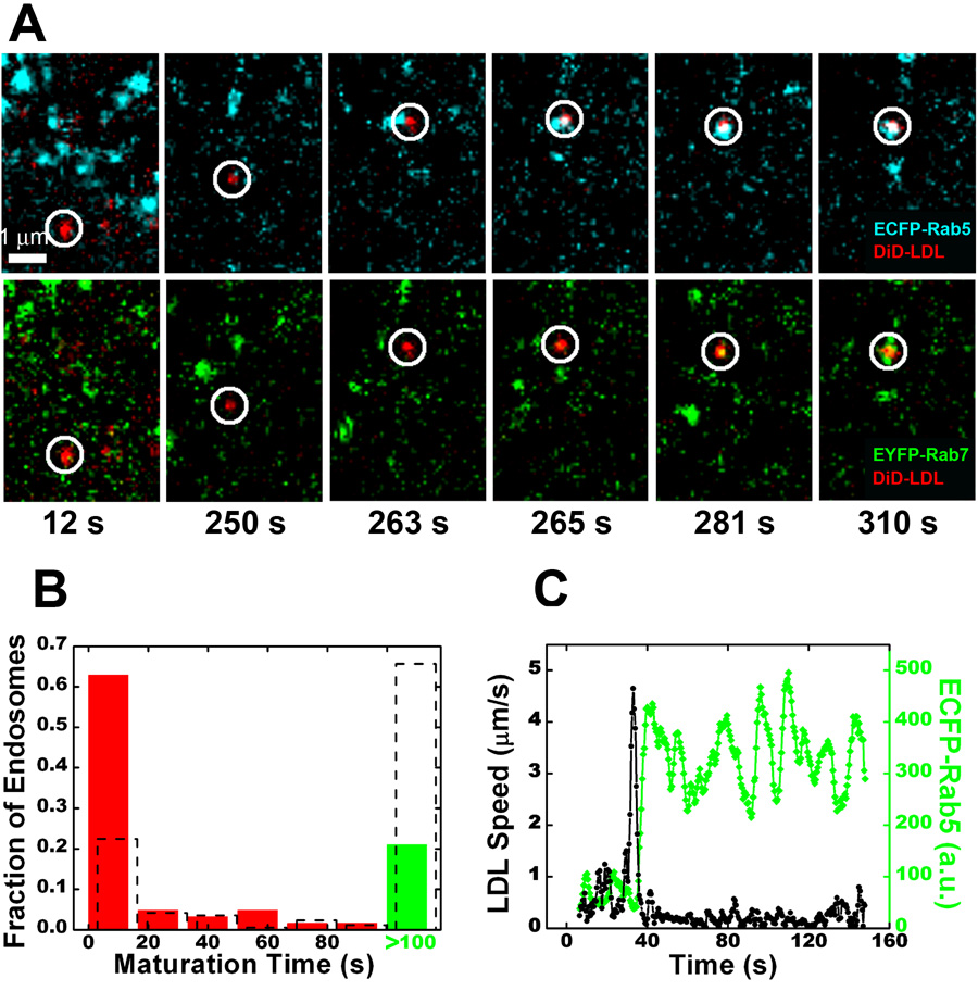
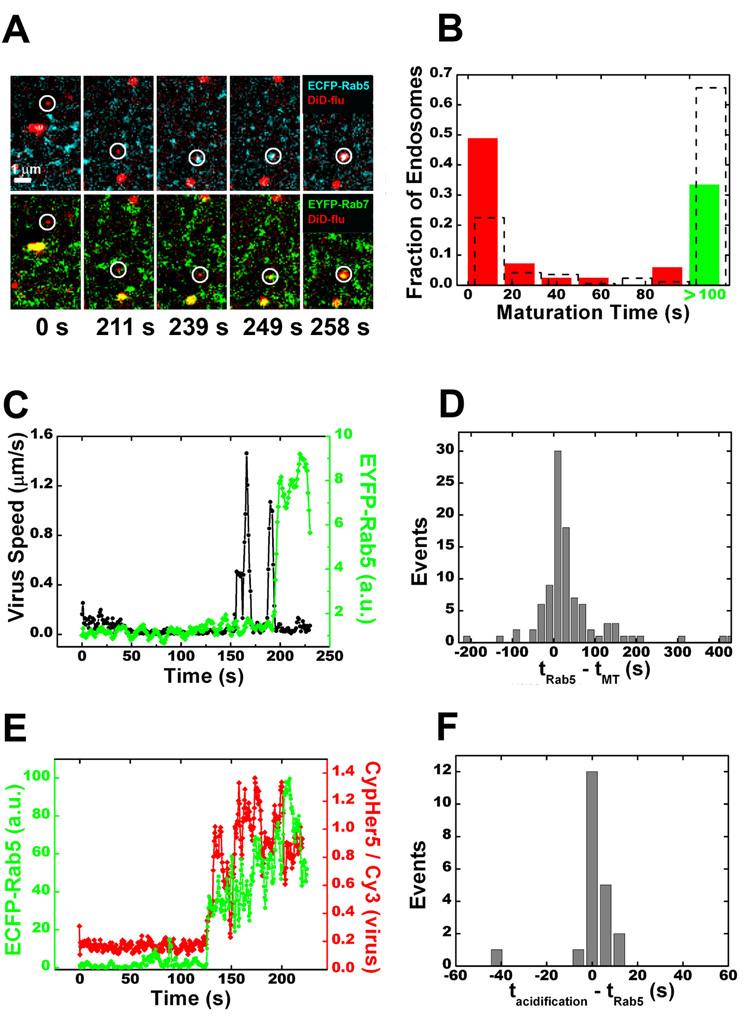
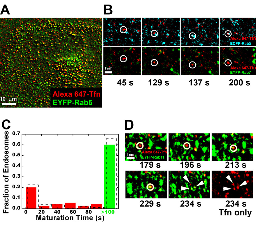
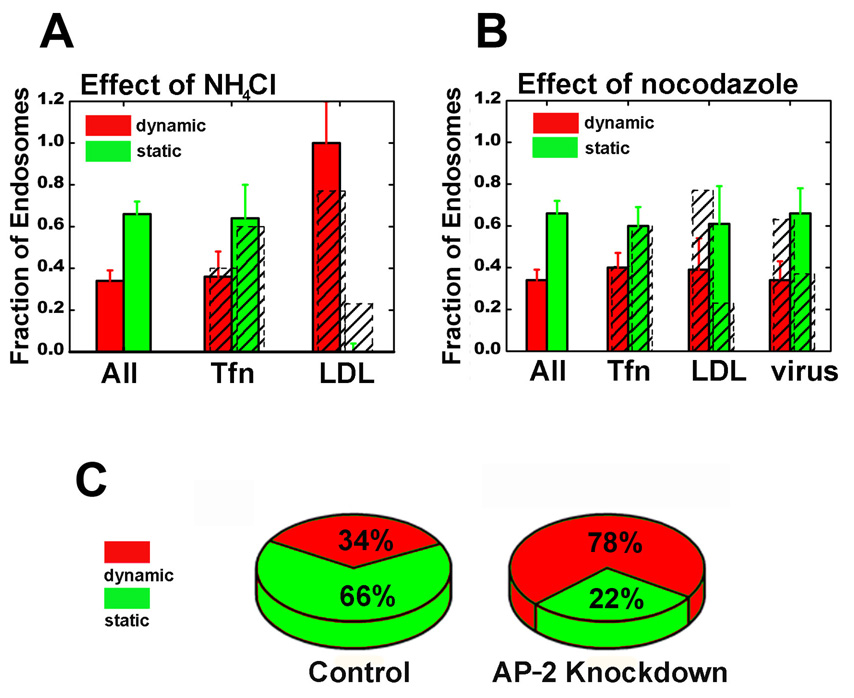
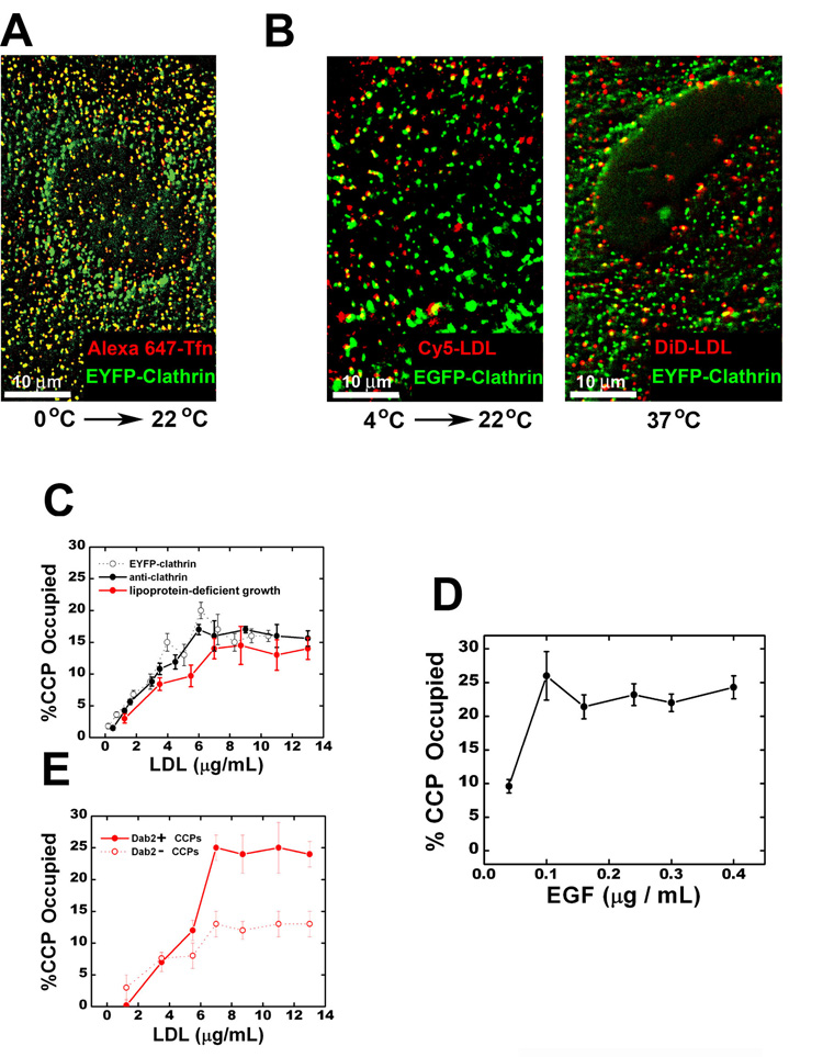
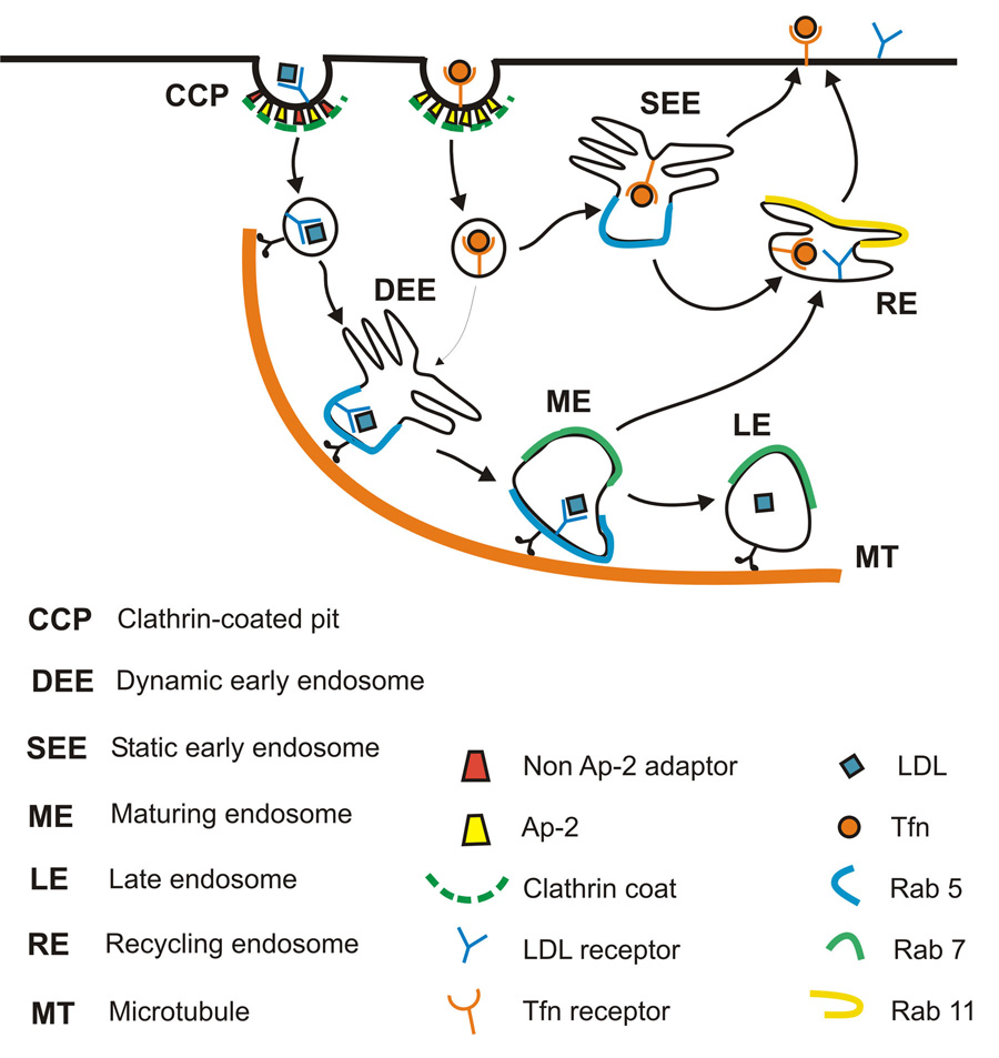
Cells rely on the correct sorting of endocytic ligands and receptors for proper function. Early endosomes have been considered as the initial sorting station where cargos for degradation separate from those for recycling. Using live-cell imaging to monitor individual endosomes and ligand particles in real time, we have discovered a sorting mechanism that takes place prior to early endosome entry. We show that early endosomes are in fact comprised of two distinct populations: a dynamic population that is highly mobile on microtubules and matures rapidly toward late endosomes and a static population that matures much more slowly. Several cargos destined for degradation are preferentially targeted to the dynamic endosomes, whereas the recycling ligand transferrin is nonselectively delivered to all early endosomes and effectively enriched in the larger, static population. This pre-early endosome sorting process begins at clathrin-coated vesicles, depends on microtubule-dependent motility, and appears to involve endocytic adaptors.


Figures

Figure 1
Figure 1
Early endosomes are comprised of two populations with distinct maturation kinetics and mobility. (A) Colocalization between Rab5 and EEA1. EEA1 was detected by immunofluorescence; Rab5 was detected by EYFP-Rab5. (B) Colocalization between Rab5, Rab7 and CI-MPR. CI-MPR was detected by immunofluorescence; Rab5 and Rab7 were detected by ECFP-Rab5 and EYFP-Rab7, respectively. The boxed region is magnified and shown in separated channels on the right. Arrowheads mark CI-MPR-positive spots that also contain Rab5 and Rab7; the arrow marks a Rab5-only endosome without CI-MPR. (C) The maturation of a Rab5-positive endosome as shown by the accumulation of Rab7 (see Movie S2). (D) The raw (black) and median-filtered (red) time-dependence of the EYFP-Rab7 intensity associated with the endosome shown in (C). (E) A histogram of the maturation time of many randomly selected Rab5-positive endosomes. Image acquisition begins at time = 0 and the maturation time is operationally defined as the time when the endosome accumulates detectable amount of Rab7. The green bar indicates endosomes with maturation time > 100 s. About 14% of the Rab5-positive endosomes already contain Rab7 at time = 0, these endosomes are included in the first bin. The inset is the maturation time histogram for the endosomes starting with only Rab5 and acquiring Rab7 within 100 seconds. The fit to a single exponential (black line) yields a time constant of 30 s. The black bar in the main histogram is the expected fraction of endosomes with maturation time > 100 s if all endosomes matured with the same single-exponential kinetics. (F) A mobility histogram of randomly selected Rab5-positive endosomes. The mobility is defined as the diameter of the smallest circle enclosing the trajectory of the endosome in 100 s. The slowly maturing endosomes (maturation time > 100 s, green) are relatively stationary compared to the rapidly maturing endosomes (maturation time ≤ 100 s, red). The shaded columns show a mobility histogram of all early endosomes in nocodazole-treated cells.
Figure 2
Figure 2
LDL is predominantly targeted to the dynamic population of early endosomes directly. (A) Selected frames from an LDL particle being trafficked to a rapidly maturing endosome (see Movie S4). The temperature was raised from 0 to 37 °C at time = 0. (B) A histogram of maturation times for endosomes receiving LDL. Overlaid is the histogram of all Rab5-positive endosomes (dashed columns). (C) Time trajectories of the velocity (black) of an LDL particle and the ECFP-Rab5 signal (green) associated with that particle. The large velocity spike indicates a long-range microtubule-dependent movement toward the perinuclear region, typically observed for endocytic ligands bound for degradation. These velocity spikes are abolished in cells treated with nocodazole.
Figure 3
Figure 3
Influenza viruses are preferentially sorted to the dynamic population of early endosomes. (A) Selected frames from a virus particle being trafficked to a rapidly maturing endosome and subsequent fusion with that endosome (see Movie S5). Fusion is indicated by a sudden increase in DiD signal due to fluorescence dequenching. Virus particles were added to the cell culture in situ and time = 0 is set for each virus when it binds to the cell. (B) A histogram of maturation times for endosomes receiving influenza. Overlaid is the histogram for all Rab5-positive endosomes (dashed columns). (C) Time trajectories of the velocity (black) of a virus particle and the Rab5 signal (green) associated with the virus. The large velocity spikes indicate long-range microtubule-dependent motion. (D) A histogram of the time lag between virus particles entering a Rab5-positive endosome (tRab5) and the onset of microtubule-dependent movement (tMT). (E) Time trajectories of the environmental acidity (red) experienced by a virus particle and the ECFP-Rab5 intensity (green) associated with the virus particle. Acidification is measured as a change in the intensity ratio between CypHer5 (a pH-sensitive dye) and Cy3 (a pH-independent dye) attached to the virus. Calibration shows that the CypHer5/Cy3 ratio changes most significantly when pH changes from neutral to pH 6 (Lakadamyali et al., 2003). (F) A histogram of the time lag between viruses joining a Rab5-positive endosome (tRab5) and the initial acidification of the virus particle (tacidification).
Figure 4
Figure 4
Transferrin is non-selectively delivered to all early endosomes. (A) Colocalization between transferrin (Tfn) and Rab5 in a cell after 5 minutes of transferrin uptake. (B) Selected frames showing a transferrin-containing vesicle being delivered to a Rab5-positive endosome that does not accumulate Rab7 within 100 s. The temperature of the sample was raised from 0 to 37 °C at time = 0. (C) The maturation time histogram of the endosomes receiving transferrin. The histogram for all endosomes (dashed columns) is overlaid for comparison. (D) Association of transferrin with Rab11-labeled recycling endosome and the subsequent tubulation and fission of the transferrin spot into several spots (arrowheads) (see Movies S6A,B).
Figure 5
Figure 5
The pre-early endosome sorting is independent of pH, but depends on microtubules and endocytic adaptor protein. (A) The effect of ammonium chloride on the balance between the dynamic (maturation time < 100 s) and static (maturation time > 100 s) endosome populations. (B) The effect of nocodazole (30 min treatment) on the balance between the two populations. In (A) and (B), colored histograms are for all endosomes, Tfn-receiving, LDL-receiving, and virus-receiving endosomes in drug-treated cells; shaded histograms are for those in untreated cells. (C) The effect of AP-2 knockdown on the balance between the two populations.
Figure 6
Figure 6
Enrichment of LDL and EGF in a subset of clathrin-coated pits (CCPs). (A) Colocalization between transferrin and clathrin. Transferrin was bound to EYFP-clathrin expressing cells at 0°C for 10 min and then imaged immediately at room temperature. (B) Colocalization between LDL and clathrin. Left panel: LDL (10 µg/ml) was bound to EYFP-clathrin expressing cells at 4°C for 10 min and then imaged immediately at room temperature. Right panel: LDL was incubated with EYFP-clathrin expressing cells at 37°C for 3 min before fixation and imaging. (C) The fraction of CCPs containing LDL as a function of the LDL concentration used. Here, LDL was incubated with cells at 37°C for 3 min. Longer LDL incubation time did not change the results. The fraction determined in EYFP-clathrin expressing cells is shown in open black symbols. To assess the effect of EYFP-clathrin expression, we determined the fraction of CCPs containing LDL in cells not expressing EYFP-clathrin (solid black symbols), with clathrin detected by immunofluorescence. Red symbols indicated results obtained in cells incubated with lipoprotein-deficient serum overnight to up-regulate LDLR expression (red symbols). (D) The fraction of CCPs containing EGF as a function of EGF concentration. The experiments were carried out similarly to (C). (E) Role of Dab2. After incubation of EGFP-clathrin expressing cells with LDL for 3 min at 37°C, cells were saponin-extracted, fixed and immunostained for Dab2. CCPs show significant colocalization with Dab2 and the quantitative extent of colocalization is cell-type dependent (Figure S6A). Solid and open red symbols, respectively, indicate the fraction of Dab2-positive and Dab2-negative CCPs containing LDL. The cells were incubated with lipoprotein-deficient serum overnight.
Figure 7
Figure 7
A proposed model for pre-early endosome sorting that differentially targets endocytic cargos into distinct populations of coated vesicles and early endosomes. Early endosomes are comprised of a dynamic population that matures quickly towards late endosomes and a relatively static population that matures much more slowly. Several cargos destined for degradation, including LDL, EGF and influenza virus, are internalized by a subpopulation(s) of clathrin-coated vesicles, which likely contain alternative adaptors in addition to AP-2. These vesicles rapidly engage microtubules and are consequently targeted to the dynamic population of early endosomes, which are also moving on microtubules. The recycling ligand transferrin is indiscriminately recruited to all clathrin-coated vesicles and thus delivered to both populations of early endosomes non-selectively, effectively being enriched in the larger, static population.

References

    1. Anderson RGW, Brown MS, Beisiegel U, Goldstein JL. Surface distribution and recycling of the low density lipoprotein receptor as visualized with antireceptor antibodies. J Cell Biol. 1982;93:523–531. - PMC - PubMed
    1. Anderson RGW, Brown MS, Goldstein JL. Role of the coated endocytic vesicle in the uptake of receptor-bound low density lipoprotein in human fibroblasts. Cell. 1977;10:351–364. - PubMed
    1. Barbero P, Bittova L, Pfeffer SR. Visualization of Rab9-mediated vesicle transport from endosomes to the trans-Golgi in living cells. J Cell Biol. 2002;156:511–518. - PMC - PubMed
    1. Bomsel M, Prydz K, Parton RG, Gruenberg J, Simons K. Endocytosis in filter-grown madin-darby canine kidney-cells. J Cell Biol. 1989;109:3243–3258. - PMC - PubMed
    1. Bonifacino JS, Lippincott-Schwartz J. Coated Proteins: shaping membrane transport. Nat Rev Mol Cell Biol. 2003;4:409–414. - PubMed

Publication types

MeSH terms