. 2006 Mar;14(3):577-87.
doi: 10.1016/j.str.2005.11.015.

Conformational flexibility in the multidrug efflux system protein AcrA

Affiliations

Conformational flexibility in the multidrug efflux system protein AcrA

Jonathan Mikolosko et al. Structure. 2006 Mar.

Abstract

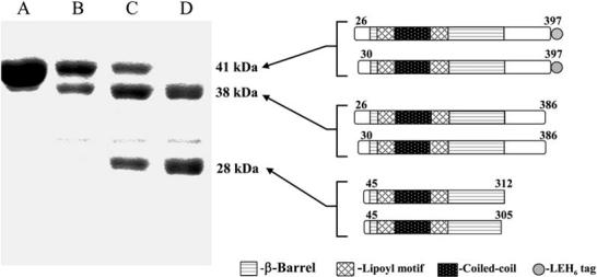
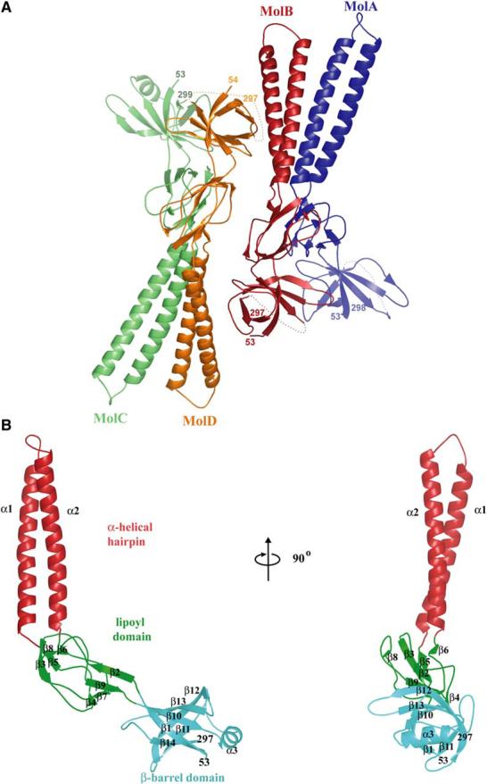
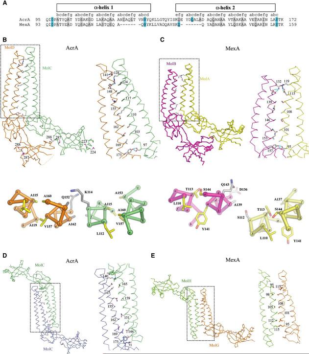
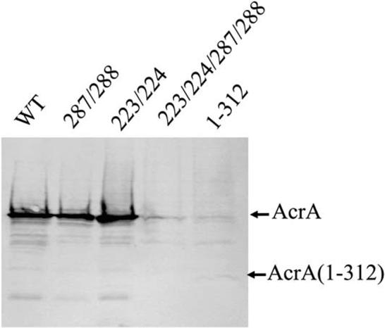
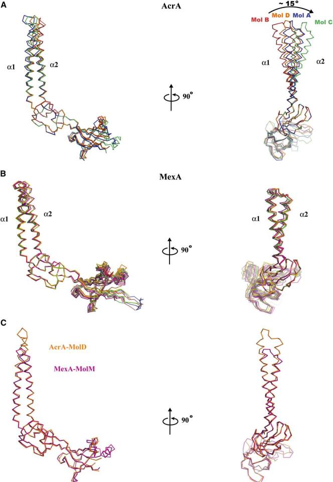
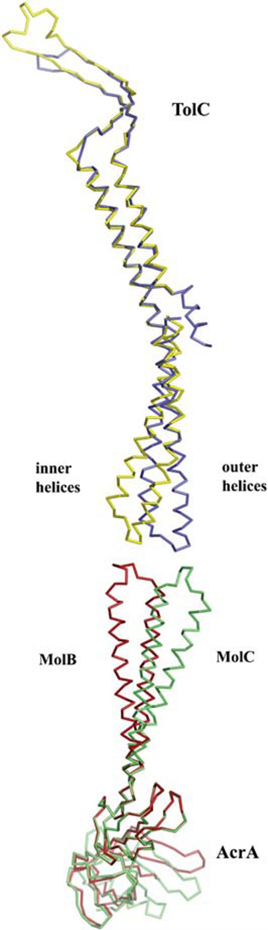
Intrinsic resistance to multiple drugs in many gram-negative bacterial pathogens is conferred by resistance nodulation cell division efflux pumps, which are composed of three essential components as typified by the extensively characterized Escherichia coli AcrA-AcrB-TolC system. The inner membrane drug:proton antiporter AcrB and the outer membrane channel TolC export chemically diverse compounds out of the bacterial cell, and require the activity of the third component, the periplasmic protein AcrA. The crystal structures of AcrB and TolC have previously been determined, and we complete the molecular picture of the efflux system by presenting the structure of a stable fragment of AcrA. The AcrA fragment resembles the elongated sickle shape of its homolog Pseudomonas aeruginosa MexA, being composed of three domains: beta-barrel, lipoyl, and alpha-helical hairpin. Notably, unsuspected conformational flexibility in the alpha-helical hairpin domain of AcrA is observed, which has potential mechanistic significance in coupling between AcrA conformations and TolC channel opening.


Figures

Figure 1
Figure 1
Domain Mapping of AcrA(26-397) by Thermolytic Digestion AcrA(26-397) was digested with thermolysin for 0, 1, 2, and 4 hr (lanes A, B, C, D, respectively) at a 50:1 (mass) substrate:protease ratio and analyzed by 12% SDS-PAGE. Molecular weights and schematics of proteolytic products are shown to the right of the corresponding fragments. Fragments were identified by N-terminal sequencing and mass spectrometry.
Figure 2
Figure 2
Structure of AcrA(45-312)-4M (A) Ribbon representation of AcrA(45-312)-4M, showing two apparent dimers per asymmetric unit in the crystal. (B) Ribbon representation of a monomer of AcrA(45-312)-4M (molecule C), with the α-helical hairpin domain in red, lipoyl domain in green, and β-barrel domain in cyan. Molecular graphics were made with PyMOL (DeLano, 2002).
Figure 3
Figure 3
Intermolecular Associations (A) Sequence alignment of the α-helical hairpins of AcrA and MexA. The corresponding heptad position is shown above the sequence with residues in the c and f positions highlighted in gray (including Ala-99, which is situated at a c position but is a b position residue according to sequence) and hydrogen bonding residues in cyan. (B) Parallel interactions of AcrA(45-312)-4M. Molecules A and B make similar dimeric contacts as molecules C and D, with the latter pair being shown here in Cα trace. Side chains are shown for residues in the c and f heptad positions as well as for residues that were substituted with methionine. The boxed region is enlarged at right, with hydrogen bonds colored in cyan. Below is a cross-section of the α-helical hairpin showing the intra- and intermolecular packing between helices. The positions of the heptad repeat are labeled a-g with side chains for the a and d heptad positions shown in yellow, and the e and f heptad positions shown in gray. (C) Parallel interactions of MexA. Molecules A and B of the 13 total molecules in the asymmetric unit of MexA are shown, with representation as in (B). (D) Antiparallel interactions of AcrA(45-312)-4M, with molecules related by crystallographic symmetry. Representation as in (B). (E) Antiparallel interactions of MexA, with molecules G and H shown and representation as in (B).
Figure 4
Figure 4
Steady-State Expression of AcrA and Mutants Western blotting analysis with anti-AcrA antibodies of total cell protein (1 μg per lane) separated on an SDS-polyacrylamide (12%) gel. AcrA was expressed along with AcrB under control of the native acrAB promoter from various derivatives of pAHisB plasmid in E. coli AG100AX (ΔacrAB::kan ΔacrEF::spe). Steady-state expression of intact AcrA(1-397) (WT), AcrA L287M/L288M, AcrA F223M/L224M, AcrA F223M/L224M/L287M/L288M, and AcrA(1-312) is shown.
Figure 5
Figure 5
Conformational Flexibility of α-Helical Hairpin (A) Comparison of four conformations of AcrA(45-312)-4M observed in the crystal, with molecules A, B, C, and D superposed on the lipoyl domain and displayed as Cα traces. The greatest difference, ∼15°, is between molecules B and C. (B) Comparison of the 13 conformations of MexA observed in its crystal, with molecules superposed on the lipoyl domain and displayed as Cα traces. (C) Superposition of molecule B of AcrA(45-312)-4M (orange) and molecule M of MexA (purple), with molecules superposed on all domains and displayed as Cα traces.
Figure 6
Figure 6
Comparison of the TolC and AcrA(45-312)-4M Coiled-Coils The magnitude of conformational change predicted for the opening of the TolC channel coincides with the flexibility observed in the AcrA α-helical hairpin domain. The inner (yellow) and outer helices (blue) of TolC are shown superposed based on the internal structural repeat (residues 16-98 and 222-316). Below these are molecules B (red) and C (green) of AcrA(45-312)-4M, as superposed on the lipoyl domain. Direct engagement of AcrA and TolC is not modeled.

References

    1. Aires JR, Nikaido H. Aminoglycosides are captured from both periplasm and cytoplasm by the AcrD multidrug efflux transporter of Escherichia coli. J. Bacteriol. 2005;187:1923–1929. - PMC - PubMed
    1. Akama H, Kanemaki M, Yoshimura M, Tsukihara T, Kashiwagi T, Yoneyama H, Narita S, Nakagawa A, Nakae T. Crystal structure of the drug discharge outer membrane protein, OprM, of Pseudomonas aeruginosa: dual modes of membrane anchoring and occluded cavity end. J. Biol. Chem. 2004;279(a):52816–52819. - PubMed
    1. Akama H, Matsuura T, Kashiwagi S, Yoneyama H, Narita S, Tsukihara T, Nakagawa A, Nakae T. Crystal structure of the membrane fusion protein, MexA, of the multidrug transporter in Pseudomonas aeruginosa. J. Biol. Chem. 2004;279(b):25939–25942. - PubMed
    1. Andersen C, Koronakis E, Bokma E, Eswaran J, Humphreys D, Hughes C, Koronakis V. Transition to the open state of the TolC periplasmic tunnel entrance. Proc. Natl. Acad. Sci. USA. 2002;99:11103–11108. - PMC - PubMed
    1. Budisa N, Steipe B, Demange P, Eckerskorn C, Kellermann J, Huber R. High-level biosynthetic substitution of methionine in proteins by its analogs 2-aminohexanoic acid, selenomethionine, telluromethionine and ethionine in Escherichia coli. Eur. J. Biochem. 1995;230:788–796. - PubMed

Publication types

MeSH terms

Associated data

LinkOut - more resources