. 2006 Apr;18(4):867-78.
doi: 10.1105/tpc.105.037135. Epub 2006 Mar 10.

Calcium-dependent protein kinase isoforms in Petunia have distinct functions in pollen tube growth, including regulating polarity

Affiliations

Calcium-dependent protein kinase isoforms in Petunia have distinct functions in pollen tube growth, including regulating polarity

Gyeong Mee Yoon et al. Plant Cell. 2006 Apr.

Abstract

Calcium is a key regulator of pollen tube growth, but little is known concerning the downstream components of the signaling pathways involved. We identified two pollen-expressed calmodulin-like domain protein kinases from Petunia inflata, CALMODULIN-LIKE DOMAIN PROTEIN KINASE1 (Pi CDPK1) and Pi CDPK2. Transient overexpression or expression of catalytically modified Pi CDPK1 disrupted pollen tube growth polarity, whereas expression of Pi CDPK2 constructs inhibited tube growth but not polarity. Pi CDPK1 exhibited plasma membrane localization most likely mediated by acylation, and we present evidence that suggests this localization is critical to the biological function of this kinase. Pi CDPK2 substantially localized to as yet unidentified internal membrane compartments, and this localization was again, at least partially, mediated by acylation. In contrast with Pi CDPK1, altering the localization of Pi CDPK2 did not noticeably alter the effect of overexpressing this isoform on pollen tube growth. Ca(2+) requirements for Pi CDPK1 activation correlated closely with Ca(2+) concentrations measured in the growth zone at the pollen tube apex. Interestingly, loss of polarity associated with overexpression of Pi CDPK1 was associated with elevated cytosolic Ca(2+) throughout the bulging tube tip, suggesting that Pi CDPK1 may participate in maintaining Ca(2+) homeostasis. These results are discussed in relation to previous models for Ca(2+) regulation of pollen tube growth.


Figures

Figure 1.
Figure 1.
Expression Patterns of Pi CDPKs. Replica RNA gel blots probed with Pi CDPK1 (A) and Pi CDPK2 (B). (C) shows ethidium bromide–stained rRNA bands as a control for loading.
Figure 2.
Figure 2.
Representative Pollen Tubes Expressing Pi CDPK1 Constructs. Images show pollen cultured in vitro for 4 h after biolistic bombardment. Paired images show GFP fluorescence micrographs on a black background (showing transformed tubes), alongside equivalent light micrographs (showing both transformed and wild-type tubes). Pollen tubes transformed with ProLat52:Pi CDPK1-GFP exhibited almost total loss of growth polarity ([A] and [B]). Tubes transformed with ProLat52:Pi CDPK1/DN-GFP appeared to gradually lose polarity as they grew, resulting in sock-like pollen tubes ([C] and [D]). Pollen transformed with ProLat52:Pi CDPK1/CA-GFP was severely inhibited in tube length and width ([E] and [F]). Control transformants expressing ProLat52:GFP are shown for comparison (G). Bars = 20 μm.
Figure 3.
Figure 3.
Representative Pollen Tubes Expressing Pi CDPK2 Constructs. Images show pollen cultured in vitro for 4 h after biolistic bombardment. Paired images show GFP fluorescence micrographs on a black background (showing transformed tubes), alongside equivalent light micrographs on a gray background (showing both transformed and wild-type tubes). Tube growth was inhibited by all Pi CDPK2 constructs, but growth polarity was not affected: ProLat52:Pi CDPK2-GFP ([A] and [B]); ProLat52:Pi CDPK2/DN-GFP ([C] and [D]); ProLat52:Pi CDPK2/CA-GFP ([E] and [F]). A control transformant expressing ProLat52:GFP is shown for comparison (G). Bars = 20 μm.
Figure 4.
Figure 4.
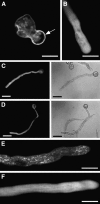
Subcellular Localization of Pi CDPKs and Its Relevance to Overexpression Phenotypes. (A) Image showing Pi CDPK1-GFP localized mainly to the cell periphery, suggesting plasma membrane localization, though some signal was also visible in the cytosol. (B) ΔN–Pi CDPK1-GFP deletion of the N-terminal acylation sites of Pi CDPK1 caused a loss of plasma membrane localization. (C) and (D) Paired images of pollen tubes expressing ΔN–Pi CDPK1-GFP growing with normal wild-type polarity in contrast with those expressing Pi CDPK1-GFP. Fluorescence micrographs show transformants only on a black background, and equivalent light micrographs show all tubes on a gray background. (E) Pi CDPK2-GFP shows a punctate distribution interpreted as localization to internal membrane compartments. (F) ΔN–Pi CDPK2-GFP fluorescence is largely cytosolic, but faint internal membrane localization is visible. (A), (B), (E), and (F) were generated using a confocal microscope (bars = 10 μm). (C) and (D) were generated using an epifluorescence microscope (bars = 20 μm).
Figure 5.
Figure 5.
Calcium Requirements for Pi CDPK1 Phosphorylation of Syntide-2. Relative kinase activity is represented as a fraction of maximal activity (2042 pmol min−1 mg−1). Results represent the mean of three separate experiments.
Figure 6.
Figure 6.
Ratiometric Ca2+ Imaging of Growing Pollen Tubes. Imaging was performed using the calcium-sensitive dye Indo-1-dextran. Cytosolic calcium concentrations are shown as indicated by the color bar. Transformed pollen tubes expressing Pi CDPK1-GFP exhibit substantial elevation of [Ca2+]i throughout the entire tip-focused gradient in the bulging tip (A) compared with both wild-type pollen tubes (C) and those expressing Pi CDPK2-GFP (B). The images shown represent approximate maximal cytosolic calcium concentration observed in the tube tips during tip growth. Bar = 10 μm.
Figure 7.
Figure 7.
Model for the Regulation of Tip Growth by Pi CDPK1. Y is a factor that in its activated form directly promotes tip growth. X is an inhibitor of Y. Z is an activator of Y. X-Y represents a complex in which X is held in an inactive state by Y. Activated Y is represented by Y*. Three regulatory steps are proposed: (1) phosphorylation of X leading to release of Y from the X-Y complex, (2) activation of free Y by Z, leading to promotion of growth, and (3) dephosphorylation of X, enabling X to interact with and inhibit Y.

References

    1. Abo-El-Saad, M., and Wu, R. (1995). A rice membrane calcium-dependent protein kinase is induced by gibberellin. Plant Physiol. 108 787–793. - PMC - PubMed
    1. Allwood, E.G., Smertenko, A.P., and Hussey, P.J. (2001). Phosphorylation of plant actin-depolymerizing factor by calmodulin-like domain protein kinase. FEBS Lett. 499 97–100. - PubMed
    1. Anil, V.S., Harmon, A.C., and Rao, K.S. (2003). Spatio-temporal accumulation and activity of calcium-dependent protein kinases during embryogenesis, seed development, and germination of sandalwood. Plant Physiol. 122 1035–1043. - PMC - PubMed
    1. Bamburg, J.R. (1999). Proteins of the ADF/cofilin family: Essential regulators of actin dynamics. Annu. Rev. Cell Biol. 15 185–230. - PubMed
    1. Battey, N.H., and Blackbourne, H.D. (1993). The control of exocytosis in plant cells. New Phytol. 125 307–338. - PubMed

Publication types

Associated data