

Increased sensitivity to cisplatin in non-small cell lung cancer cell lines after FHIT gene transfer
- PMID: 16533421
- PMCID: PMC1584285
- DOI: 10.1593/neo.05517
Increased sensitivity to cisplatin in non-small cell lung cancer cell lines after FHIT gene transfer
Abstract
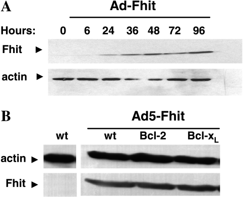
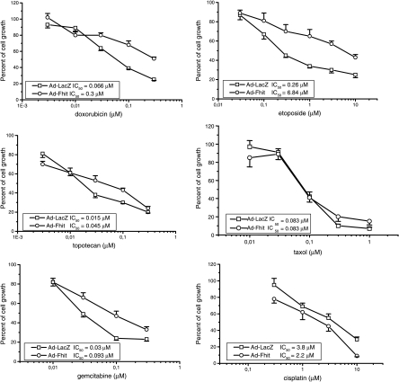
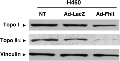
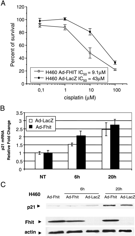
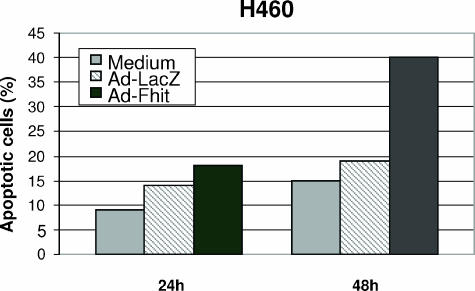
To evaluate the relevance of fragile histidine triad (FHIT) status in relation to drug treatment, we analyzed the sensitivity of the Fhit-negative non-small cell lung cancer (NSCLC) cell line NCI-H460 to different drugs, after treatment with an adenoviral vector expressing the FHIT transgene. Expression of Fhit resulted in reduced sensitivity to etoposide, doxorubicin, and topotecan. This feature was associated with Fhit-induced downregulation of DNA topoisomerases I and II. In contrast, expression of Fhit did not modulate sensitivity to Taxol, but produced a slight increase in sensitivity to cisplatin, as shown by colony-forming assays. Analysis of apoptosis revealed that, after cisplatin exposure, the number of apoptotic cells was two-fold higher in Fhit-expressing H460 cells. Moreover, it appeared that wildtype p53 was required for sensitization to cisplatin because the effect was marginal in A549 and Calu-1 cells, where the p53 pathway is altered and simultaneous restoration of p53 and Fhit in Calu-1 cells increased cisplatin sensitivity. Fhit could also partially restore sensitivity to cisplatin in Bcl-2- and Bcl-x(L)-overexpressing H460 cells that are normally resistant to this drug. Our results support the possible relevance of FHIT in cisplatin-based chemotherapy as well as in the reversal of drug resistance in NSCLC.
Figures


Similar articles
-
The apoptotic pathway triggered by the Fhit protein in lung cancer cell lines is not affected by Bcl-2 or Bcl-x(L) overexpression.Oncogene. 2004 Dec 2;23(56):9102-10. doi: 10.1038/sj.onc.1208142. Oncogene. 2004. PMID: 15489891
-
Dose-dependent effect of FHIT-inducible expression in Calu-1 lung cancer cell line.Oncogene. 2004 Nov 4;23(52):8439-46. doi: 10.1038/sj.onc.1207847. Oncogene. 2004. PMID: 15361849
-
The tumor-suppressor gene FHIT is involved in the regulation of apoptosis and in cell cycle control.Proc Natl Acad Sci U S A. 1999 Jul 20;96(15):8489-92. doi: 10.1073/pnas.96.15.8489. Proc Natl Acad Sci U S A. 1999. PMID: 10411902 Free PMC article.
-
FHIT loss confers cisplatin resistance in lung cancer via the AKT/NF-κB/Slug-mediated PUMA reduction.Oncogene. 2015 May 7;34(19):2505-15. doi: 10.1038/onc.2014.184. Epub 2014 Jul 7. Oncogene. 2015. PMID: 24998847
-
The clinicopathological significance of FHIT hypermethylation in non-small cell lung cancer, a meta-analysis and literature review.Sci Rep. 2016 Jan 22;6:19303. doi: 10.1038/srep19303. Sci Rep. 2016. PMID: 26796853 Free PMC article. Review.
Cited by
-
CDKN1A upregulation and cisplatin‑pemetrexed resistance in non‑small cell lung cancer cells.Int J Oncol. 2020 Jun;56(6):1574-1584. doi: 10.3892/ijo.2020.5024. Epub 2020 Mar 24. Int J Oncol. 2020. PMID: 32236605 Free PMC article.
-
The antitumour efficacy of hesperidin vs. cisplatin against non-small lung cancer cells A549 and H460 via targeting the miR-34a/PD-L1/NF-κB signalling pathway.Contemp Oncol (Pozn). 2024;28(2):130-148. doi: 10.5114/wo.2024.141648. Epub 2024 Jul 19. Contemp Oncol (Pozn). 2024. PMID: 39421711 Free PMC article.
-
Neoplasia: the second decade.Neoplasia. 2008 Dec;10(12):1314-24. doi: 10.1593/neo.81372. Neoplasia. 2008. PMID: 19048110 Free PMC article.
-
IL-6 signaling contributes to cisplatin resistance in non-small cell lung cancer via the up-regulation of anti-apoptotic and DNA repair associated molecules.Oncotarget. 2015 Sep 29;6(29):27651-60. doi: 10.18632/oncotarget.4753. Oncotarget. 2015. PMID: 26313152 Free PMC article.
-
Transcutaneous electrical acupoint stimulation (TEAS) ameliorates chemotherapy-induced bone marrow suppression in lung cancer patients.J Thorac Dis. 2017 Mar;9(3):809-817. doi: 10.21037/jtd.2017.03.12. J Thorac Dis. 2017. PMID: 28449490 Free PMC article.
References
-
- Ferreira CG, Huisman C, Giaccone G. Novel approaches to the treatment of non-small cell lung cancer. Crit Rev Oncol Hematol. 2002;41:57–77. - PubMed
-
- Nishio K, Nakamura T, Koh Y, Suzuki T, Fukumoto H, Saijo N. Drug resistance in lung cancer. Curr Opin Oncol. 1999;11:109–115. - PubMed
-
- Wang G, Reed E, Li QQ. Molecular basis of cellular response to cisplatin chemotherapy in non-small cell lung cancer. Oncol Rep. 2004;12:955–965. (Review) - PubMed
-
- Schiller JH, Harrington D, Belani CP, Langer C, Sandler A, Krook J, Zhu J, Johnson DH. Comparison of four chemotherapy regimens for advanced non-small-cell lung cancer. N Engl J Med. 2002;346:92–98. - PubMed
-
- Sozzi G, Pastorino U, Moiraghi L, Tagliabue E, Pezzella F, Ghirelli C, Tornielli S, Sard L, Huebner K, Pierotti MA, et al. Loss of FHIT function in lung cancer and preinvasive bronchial lesions. Cancer Res. 1998;58:5032–5037. - PubMed
MeSH terms
Substances
LinkOut - more resources
Full Text Sources
Other Literature Sources
Medical
Research Materials
Miscellaneous