. 2006 Mar 1;12(5):1431-40.
doi: 10.1158/1078-0432.CCR-05-1806.

Kinetics of metastatic breast cancer cell trafficking in bone

Affiliations

Kinetics of metastatic breast cancer cell trafficking in bone

Pushkar A Phadke et al. Clin Cancer Res. .

Abstract

Purpose: In vivo studies have focused on the latter stages of the bone metastatic process (osteolysis), whereas little is known about earlier events, e.g., arrival, localization, and initial colonization. Defining these initial steps may potentially identify the critical points susceptible to therapeutic intervention.

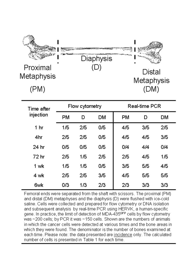
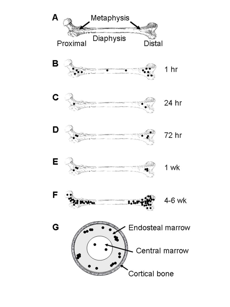
Experimental design: MDA-MB-435 human breast cancer cells engineered with green fluorescent protein were injected into the cardiac left ventricle of athymic mice. Femurs were analyzed by fluorescence microscopy, immunohistochemistry, real-time PCR, flow cytometry, and histomorphometry at times ranging from 1 hour to 6 weeks.

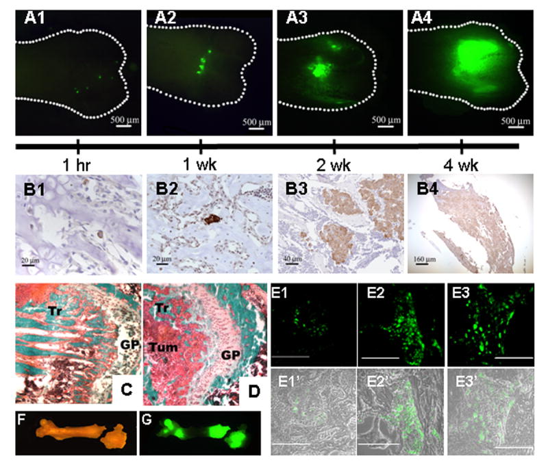
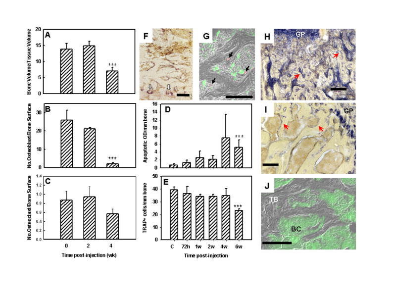
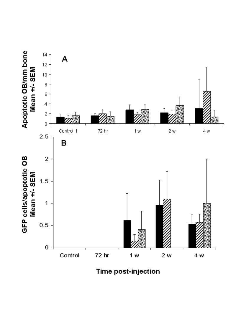
Results: Single cells were found in distal metaphyses at 1 hour postinjection and remained as single cells up to 72 hours. Diaphyseal arrest occurred rarely and few cells remained there after 24 hours. At 1 week, numerous foci (2-10 cells) were observed, mostly adjacent to osteoblast-like cells. By 2 weeks, fewer but larger foci (> or =50 cells) were seen. Most bones had a single large mass at 4 weeks (originating from a colony or coalescing foci) which extended into the diaphysis by 4 to 6 weeks. Little change (<20%) in osteoblast or osteoclast numbers was observed at 2 weeks, but at 4 to 6 weeks, osteoblasts were dramatically reduced (8% of control), whereas osteoclasts were reduced modestly (to approximately 60% of control).

Conclusions: Early arrest in metaphysis and minimal retention in diaphysis highlight the importance of the local milieu in determining metastatic potential. These results extend the Seed and Soil hypothesis by demonstrating both intertissue and intratissue differences governing metastatic location. Ours is the first in vivo evidence that tumor cells influence not only osteoclasts, as widely believed, but also eliminate functional osteoblasts, thereby restructuring the bone microenvironment to favor osteolysis. The data may also explain why patients receiving bisphosphonates fail to heal bone despite inhibiting resorption, implying that concurrent strategies that restore osteoblast function are needed to effectively treat osteolytic bone metastases.


Figures

Fig 1
Fig 1
The kinetics of MDA-435GFP metastatic growth in the femur following intracardiac injection. Whole femurs were dissected and fluorescent foci were visualized in the intact bones using a fluorescent stereomicroscope. Panel A, fluorescent foci were observed, mainly in the distal end of femurs, as shown at 1 hr (A1), 1 wk (A2), 2 wk (A3), and 4 wk (A4). Panel B, MDA-435GFP cells were detected by anti-GFP immunohistochemistry (cells stain brown) in femurs at 1 hr (B1; note: single cell), 1 wk (B2; note: clusters of 2–3 cells), 2 wk (B3), and 4 wk (B4). With time, the number of fluorescent foci decreased as the size increased. Independent tumor deposits often coalesced. Panels C and D, representative images of distal ends of femur stained with Goldner’s trichrome stain (Panel C – normal bone, Panel D – 4 wk). The amount of trabecular bone (Tr, stained teal) is significantly lower in bone containing tumor cells, reflective of osteolytic degradation. Tumor cells (Tum) infiltrating the metaphyseal area near epiphyseal growth plate (GP) are labeled for reference. Panel E shows fluorescent tumor cell foci in trabecular bone in paraffin-embedded sections at 2 wk (Panel E1), 4 wk (Panel E2) and 6 wk (Panel E3) post-injection. Magnification line indicates 100 μm. Panels E1′, E2′ and E3′ are composite of fluorescent and phase images. Representative bright field (Panel F) and fluorescent (Panel G) images of a mouse femur at 4 wk show two large metastatic foci, one at each end. The distal end shows an iatrogenic fracture, presumably due to weakness caused by tumor cell-induced osteolysis.
Fig. 2
Fig. 2
Detection of MDA-435GFP metastatic cells by flow cytometry or real-time, quantitative PCR in the metaphyseal and diaphyseal ends of the femur at various times following intracardiac inoculation.
Fig. 3
Fig. 3
MDA-435GFP breast cancer cells diminished osteoblast (OB) and osteoclast (OC) numbers in colonized bone as evaluated by quantitative bone histomorphometry, immunohistochemistry and fluorescent microscopy. Panels A–C, histomorphometric analyses. (A, Bone volume to tissue volume; B, Number of OB per bone surface; C, Number of OC per bone surface). Panel D, the number of apoptotic OB (TUNEL positive) per linear bone surface at times following inoculation of tumor cells. Panel E, the number of OC (staining for tartrate-resistant acid phosphatase, TRAP) per linear bone surface at times following inoculation of tumor cells. Panels A–E, *** indicates significantly different (p ≤ 0.05 ) from normal bone. Panel F, representative image of OC staining for TRAP (red stain highlighted with white arrows) taken from a section of femur 2 wk after tumor cell inoculation. Panel G, merged photomicrograph of MDA-435GFP tumor cells (green) surrounding apoptotic OB (red by TUNEL using Cy-5 probe) taken from a femur 6 wk following inoculation. Panels H, I, J, cryosections from a femur taken 4 wk after tumor inoculation. H,I, stained for alkaline phosphatase activity (blue staining highlighted by red arrows) indicative of OB; J, merged fluorescent and phase images showing trabecular bone (TB) surrounded by MDA-435GFP cells (BC). Alkaline phosphatase activity was greatly diminished in the trabecular bone of tumor-bearing femurs but was still present in the growth plate (GP). Bars = 100 μm.
Fig. 4
Fig. 4
The presence of metastatic breast cancer cells in close proximity to apoptotic osteoblasts increased with time following inoculation of MDA-435GFP. Panel A, apoptotic OB, detected by TUNEL, were counted in proximal and distal ends of paraffin sections of femur at times following tumor cell inoculation. Panel B, number of MDA-435GFP cells within a 50 μm radius of each apoptotic OB. Shown are the averages from three femurs per time. Proximal femur (hatched); distal femur (stippled); average over femur (solid). Apoptotic osteoblasts in the diaphyses were extremely rare.
Fig. 5
Fig. 5
Schematic diagram depicting colonization of the femur by MDA-435GFP cells. A normal femur is diagramed and labeled for reference (Panel A). Single cells (•) arrive in the bone marrow within 1 hr after intracardiac injection (Panel B), with a distribution proportionate to the relative blood flow to regions of the bone. Most cells arresting in the bone are cleared within 24 hr (Panel C). Of those remaining, the vast majority are still single cells but all are located in the metaphyses. A fraction of the surviving cells begin to proliferate by 72 hr (Panel D) with little change in the number of foci, or size of tumor cell clusters, at one week post-inoculation (Panel E). The lesions progressively grow in size so that by 4-6 wk, the mass of the metastases is large and the number of independently seeded cells indiscernible because the foci have coalesced. Despite not seeding and remaining in the diaphysis, metastases extend into the bone shaft as the lesions grow (Panel F). Flushing of bone marrow in established metastases as depicted in Figure 2, revealed that most of the tumor cells are found in endosteal marrow (~90%) or in the central marrow (~10%), but never in the cortical bone of the diaphysis, as depicted in a cross-sectional view (Panel G).

Similar articles

Cited by

References

    1. Body JJ. Metastatic bone disease: clinical and therapeutic aspects. Bone. 1992;13 (Suppl 1):S57–S62. - PubMed
    1. Coleman RE. Skeletal complications of malignancy. Cancer. 1997;80:1588–94. - PubMed
    1. Coleman RE, Rubens RD. The clinical course of bone metastases from breast cancer. Br J Cancer. 1987;55:61–66. - PMC - PubMed
    1. Mundy GR. Metastasis: Metastasis to bone: causes, consequences and therapeutic opportunities. Nature Rev Cancer. 2002;2:584–93. - PubMed
    1. Lelekakis M, Moseley JM, Martin TJ, et al. A novel orthotopic model of breast cancer metastasis to bone. Clin Exptl Metastasis. 1999;17:163–70. - PubMed

Publication types

Substances