. 2006 Jun 1;90(11):3983-92.
doi: 10.1529/biophysj.105.076406. Epub 2006 Mar 13.

Folding, misfolding, and amyloid protofibril formation of WW domain FBP28

Affiliations

Folding, misfolding, and amyloid protofibril formation of WW domain FBP28

Yuguang Mu et al. Biophys J. .

Abstract

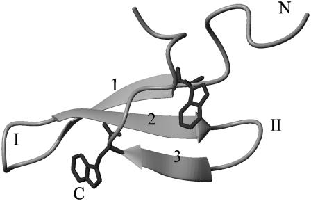
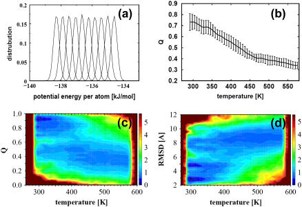
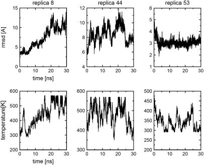
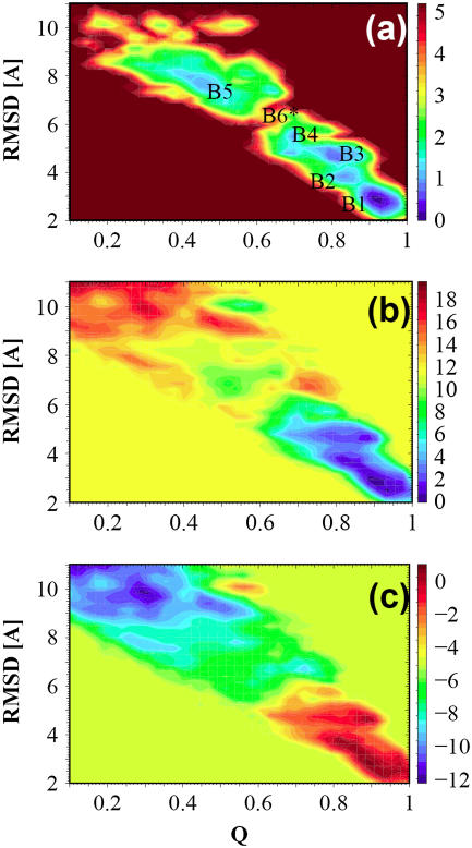
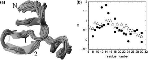
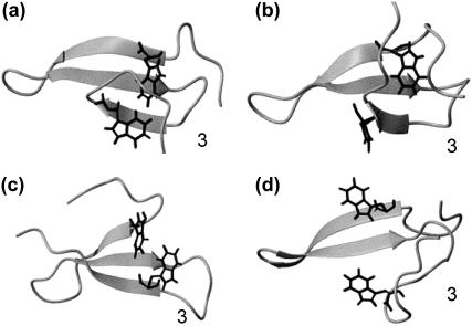
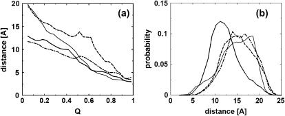
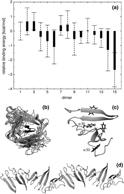
We study the folding mechanism of a triple beta-strand WW domain from the Formin binding protein 28 (FBP28) at atomic resolution with explicit water model using replica exchange molecular dynamics computer simulations. Extended sampling over a wide range of temperatures to obtain the free energy, enthalpy, and entropy surfaces as a function of structural coordinates has been performed. Simulations were started from different configurations covering the folded and unfolded states. In the free energy landscape a transition state is identified and its structures and -values are compared with experimental data from a homologous protein, the prolyl-isomerase Pin1 WW domain. A stable intermediate state is found to accumulate during the simulation characterized by the carboxyl-terminal beta-strand 3 having misregistered hydrogen bonds and where the structural heterogeneity is due to nonnative turn II formation. Furthermore, the aggregation behavior of the FBP28 WW domain may be related to one such misfolded structure, which has a much lower free energy of dimer formation than that of the native dimer. Based on the misfolded dimer, aggregation to form protofibril structure is discussed.


Figures

FIGURE 1
FIGURE 1
Experimental folded structure from the Protein Data Bank 1E0L model 1(8). N-, C-terminal, strands 1, 2, and 3, and turn I and II are labeled. The side chains of two conserved tryptophan residues (8 and 30) are shown explicitly.
FIGURE 2
FIGURE 2
(a) Distributions of potential energy per atom of the first 10 replicas whose temperatures range from 290 K to 340 K. (b) The average fraction of native contacts formed, Q, as a function of the temperature. The error bars are calculated by five-block averages. (c) Relative population of sampled conformations as a function of Q and temperature. (d) Relative population of sampled conformations as a function of RMSD and temperature. The coloring scheme in c and d is based on the free energy-like quantity explained in the main text.
FIGURE 3
FIGURE 3
Trajectories of RMSD and temperature as a function of time, sampled by replicas 8, 44, and 53.
FIGURE 4
FIGURE 4
(a) Free energy landscape, ΔG, as a function of Q and RMSD for temperature 300 K. (b) The enthalpy contribution ΔH and (c) entropy contribution, −TΔS, T = 300 K. All coloring labels are in units of kcal/mol.
FIGURE 5
FIGURE 5
(a) Ensemble of structures of the transition state. (b) Comparison of the calculated Φ-values (▵) with the experimental determined Φ-values for the Pin1 WW domain, solid circles for side chain mutation results, and solid squares for amide-to-ester mutation results (11).
FIGURE 6
FIGURE 6
Representative structures of the misfolded FBP28 WW domain, a, b, c, and d. Strand 3 makes misregistered hydrogen bonds with strand 2, with one, two, and three amino acids shifted in a, b, and c, respectively. In the misfolded structure (d), the strand 3 makes a U-turn.
FIGURE 7
FIGURE 7
(a) Distances as a function of Q averaged at the transition temperature T = 375 K. Solid, dotted, dashed, and dotted-dashed lines represent D1: the distance between amide nitrogen of residue 9 and carbonyl oxygen of residue 21 (9:N–21:O); D4, 11:O—19:N; D5, 20:N—29:O; and D8, 22:O—27:N. (b) The distribution of the probability of head-to-tail distance for peptide segments taken from FBP28 turn I (solid line), FBP28 turn II (dotted line), Pin1 turn II (dashed line), and YAP turn II (dotted-dashed line).
FIGURE 8
FIGURE 8
(a) Relative binding energies of dimers, calculated by averaging the lowest 20 docking energies from 100 docked configurations for each dimer. The labels indicate different dimers with 1–15 representing the dimer of N-N, N-a, N-b, N-c, N-d, a-a, a-b, a-c, a-d, b-b, b-c, b-d, c-c, c-d, and d-d, respectively, of which N symbolizes the native state. a, b, c, and d refer to the misfolded structures a, b, c, and d shown in Fig. 6. (b) Superposition of 20 misfolded-type-d-formed (Fig. 6) homodimer structures, which have the lowest binding energy; the target monomer is shown in the middle with dark color. (c) One homodimer structure formed by the misfolded type d in Fig. 6. Four tryptophan residues are shown, of which the two W30 are labeled. (d) A protofiber chain consisting of 10 monomers formed in the same way as the dimer does in c.

References

    1. Fersht, A. 1998. Enzyme Structure, Mechanism & Protein Folding, 3rd ed. W. H. Freeman, New York.
    1. Ferrara, P., and A. Caflisch. 2000. Folding simulations of a three-stranded antiparallel beta-sheet peptide. Proc. Natl. Acad. Sci. USA. 97:10780–10785. - PMC - PubMed
    1. Kubelka, J., J. Hofrichter, and W. A. Eaton. 2004. The protein folding ‘speed limit’. Curr. Opin. Struct. Biol. 14:76–88. - PubMed
    1. Zhou, Y., and M. Karplus. 1999. Interpreting the folding kinetics of helical proteins. Nature. 401:400–403. - PubMed
    1. Munoz, V., P. A. Thompson, J. Hofrichter, and W. A. Eaton. 1997. Folding dynamics and mechanism of beta-hairpin fromation. Nature. 390:196–199. - PubMed

Publication types