. 2006 Jun 1;90(11):3966-82.
doi: 10.1529/biophysj.105.078691. Epub 2006 Mar 13.

Model of chromosome motility in Drosophila embryos: adaptation of a general mechanism for rapid mitosis

Affiliations

Model of chromosome motility in Drosophila embryos: adaptation of a general mechanism for rapid mitosis

G Civelekoglu-Scholey et al. Biophys J. .

Abstract

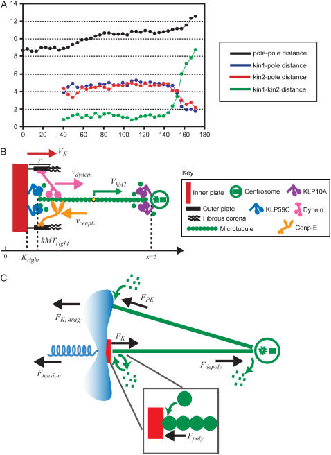
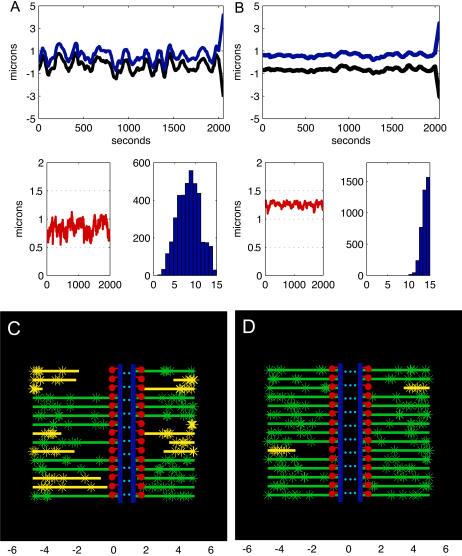
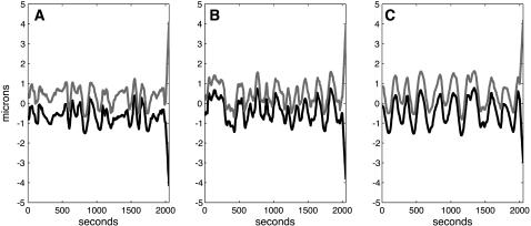
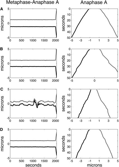
During mitosis, ensembles of dynamic MTs and motors exert forces that coordinate chromosome segregation. Typically, chromosomes align at the metaphase spindle equator where they oscillate along the pole-pole axis before disjoining and moving poleward during anaphase A, but spindles in different cell types display differences in MT dynamicity, in the amplitude of chromosome oscillations and in rates of chromatid-to-pole motion. Drosophila embryonic mitotic spindles, for example, display remarkably dynamic MTs, barely detectable metaphase chromosome oscillations, and a rapid rate of "flux-pacman-dependent" anaphase chromatid-to-pole motility. Here we develop a force-balance model that describes Drosophila embryo chromosome motility in terms of a balance of forces acting on kinetochores and kMTs that is generated by multiple polymer ratchets and mitotic motors coupled to tension-dependent kMT dynamics. The model shows that i), multiple MTs displaying high dynamic instability can drive steady and rapid chromosome motion; ii), chromosome motility during metaphase and anaphase A can be described by a single mechanism; iii), high kinetochore dynein activity is deployed to dampen metaphase oscillations, to augment the basic flux-pacman mechanism, and to drive rapid anaphase A; iv), modulation of the MT rescue frequency by the kinetochore-associated kinesin-13 depolymerase promotes metaphase chromosome oscillations; and v), this basic mechanism can be adapted to a broad range of spindles.


Figures

FIGURE 1
FIGURE 1
Metaphase and anaphase A chromatid motility in Drosophila embryos; qualitative and force-balance model. (A) Dynamics of spindle poles and chromatids in Drosophila embryos. During metaphase (∼80–135 s), the chromatids remain at the spindle equator, and do not exhibit oscillations between the spindle poles as observed in some other organisms. During anaphase A (∼135–175 s), chromatids move steadily and rapidly toward the spindle poles, which are held at constant spacing at ∼10 μm (14,18). (B) Kinetochore-MT interface in Drosophila embryos, adapted from Maiato (5) and Rogers et al. (18). A kinetochore inner (red) and outer plate (black) along with the fibrous corona (black), a dynein (pink), and a cenpE (orange) motor generating antagonistic forces at the kinetochore, the KLP59C motors (blue) depolymerizing MT's (green) plus end inserted into the kinetochore, a centrosome (green circle), and the KLP10A motors (purple) depolymerizing the minus end of the MT at the spindle pole are shown. The direction of the velocities of motors, kinetochore and MT, and the position of the spindle equator (x = 0), the kinetochore plate, the plus end of the kMT, and the right spindle pole (x = 5) are indicated. (C) Force-balance model. Forces acting on the kinetochore and the kMT are shown. For simplicity, only a single kMT is shown bound to the kinetochore, and only a single spindle MT impinging on the chromosome arm generating polar ejection forces is shown.
FIGURE 2
FIGURE 2
Metaphase and anaphase A chromatid dynamics is sensitive to MT turnover. (A) (Upper plot) Positions of sister kinetochores versus time during metaphase (initial 2000 s) and anaphase A (from 2000 to 2050 s) in a spindle where MTs turn over very rapidly. The left kinetochore (black) is tethered to the left spindle pole located at −5 μm from spindle equator and its sister kinetochore (blue) is tethered to the right spindle pole (located at 5 μm from spindle equator) throughout the duration of the metaphase and anaphase A. The initial conditions are as described in the Appendix, and simulations are run for 2000 s to stabilize before the recording. The MT dynamic parameters are vg = vs = 0.25 μm s−1; fres = 0.1 s−1; fcat = 0.06 s−1; nd = 15 μm−1; the kinetochores have 15 MT binding sites, and all other parameters are as shown in Table 1. During metaphase (initial 2000 s), the sister chromatids oscillate around the spindle equator, the mean duration of poleward or antipoleward oscillations is ∼50–100 s, and the distance traveled during a poleward or anti-poleward excursion is ∼0.5–2 μm, whereas the MTs flux toward the spindle poles at rate vflux = 0.048 ± 0.015 μm s−1 and polymerize/depolymerize at their plus ends near the kinetochores (see movie 1 in the Supplementary Material for the dynamics of MTs). After the dissolution of the cohesin links between the sisters (at 2000th s), during anaphase A (from 2000 to 2050 s), the chromatids move along the kt-fibers steadily at a rate vA ∼ 0.065 μm s−1 toward their respective poles, despite the highly dynamic nature of the MTs they are attached to. Note that the MTs continue to flux toward the poles (vflux = 0.049 ± 0.016 μm s−1) and polymerize/depolymerize at their plus ends during anaphase A, according to the same rules as in metaphase; however, the sister kinetochore tension and polar ejection forces being set to zero no longer contribute to the force on the kinetochore, which alters the transition frequencies. (Lower left plot) Distance between the sister kinetochores during metaphase, the rest length of the cohesin link between the sisters is 0.5 μm; kinetochores thus remain almost always under tension, at ∼0.9 μm average distance from one another. (Lower right plot) Histogram of number of MTs attached to kinetochores during metaphase, value 8 ± 3 (data from left and right kinetochores were pooled together since there was no significant difference in the separately calculated mean values and standard deviations). (B) (Upper plot) Positions of sister kinetochores versus time during metaphase (initial 2000 s) and anaphase A (from 2000 to 2050 s) in a spindle where MTs turn over slowly. The left (black) and right (blue) kinetochore and spindle poles, and the initial conditions and parameters are as described in A, except fres = 0.02 s−1; fcat = 0.0012 s−1. During metaphase (initial 2000 s), the sister chromatids remain stably around the spindle equator, jiggle only very little, whereas the MTs flux toward the spindle poles at rate vflux = 0.047 ± 0.0061 μm s−1 and polymerize/depolymerize at their plus ends near the kinetochores (see movie 2 for the dynamics of MTs). During anaphase A (from 2000 to 2050 s), the chromatids move along the kt-fibers steadily at a rate vA ∼ 0.055 μm s−1 toward their respective poles, whereas MTs continue to flux toward the poles at the rate vflux = 0.05 ± 0.008 μm s−1. (Lower left plot) Distance between the sister kinetochores during metaphase, the rest length of the cohesin link between the sisters is 0.5 μm; kinetochores thus remain always under tension, at ∼1.3 μm average distance from one another. (Lower right plot) Histogram of number of MTs attached to kinetochores during metaphase, value 14 ± 1 (a higher value than in A). (C) A snapshot from the simulation (movie 1) of kinetochore motility in a spindle, where MTs turn over rapidly as in A, is shown. The left and right kinetochore plates are shown in blue, the cohesin bonds are blue dotted lines between the kinetochores, and 15 MTs that transiently bind to the kinetochores (green and yellow lines) are shown. The left and the right spindle poles are located at −5 and 5, respectively, along the horizontal axis. All MT minus ends terminate near the spindle poles, whereas MTs undergo poleward flux and MT plus ends undergo dynamic instability. The MTs whose plus ends are currently interacting with the kinetochore are shown in green; others are shown in yellow. The green/yellow stars on MTs are fiduciary marks, representing the tubulin speckles on the MTs. The red lines and dots represent the fibrous corona and the outer kinetochore structure where the MT plus ends are inserted in and attach to the kinetochore. (D) A snapshot from the simulation (movie 2) of kinetochore motility in a spindle, where MTs turn over slowly as in B, is shown. Definitions of the lines and colors are as in C. Note that more MTs are bound to the kinetochores compared with C, and the kinetochores are positioned at the spindle equator.
FIGURE 3
FIGURE 3
Kinetochore size does not affect metaphase oscillations but affects anaphase A rates. Positions of the sister chromatids versus time during metaphase (initial 2000 s) and anaphase A (from 2000 to 2050 s) in spindles, where MTs turn over rapidly and the kinetochores have 7 (A), 15 (B), and 30 (C) MT binding sites, are shown. In all three figures, positions of the left and right kinetochore are shown in black and gray, respectively. The positions of the spindle poles, and the initial conditions and all parameters except the number of MT binding sites at the kinetochores, are as described in Fig. 2A. In all three cases, during metaphase (initial 2000s) kinetochores switch between poleward and antipoleward movements and thus exhibit directional instability, although the excursions become smoother and more regular as kinetochore-MT binding site increases. During Anaphase A (2000–2050 s), the kt to pole rate increases slightly with decreasing number of kinetochore MT binding sites: vA ∼ 0.075, 0.07, and 0.065 μm s−1, for A, B, and C, respectively. The average number of MTs bound to the kinetochore is 5 ± 1, 8 ± 3, and 20 ± 5 for the kinetochores in A–C, respectively.
FIGURE 4
FIGURE 4
Dynamics of kinetochores during metaphase and anaphase A is sensitive to dynein and KLP59C activity at the kinetochores. In all the figures, the positions of the left and right kinetochores are shown in black and gray, and the left and right spindle poles are at −5 and 5, respectively. (A) High dynein activity level at the kinetochores damps the metaphase oscillations and increases anaphase A rates. Positions of sister chromatids versus time during metaphase (initial 2000 s in the left panel) and anaphase A (in the right panel, the last 50 s are blown up from the left panel) in a spindle where MTs turn over rapidly is shown in spindles with high levels of dynein activity at the kinetochores with 15 MT binding sites. In both panels, the initial conditions and all parameters, except for nd = 30 and ɛ = 50 pN μm−1, are as in Fig. 2 A. Oscillations of sister chromatids around the spindle equator during metaphase (initial 2000 s) are absent, and anaphase A (from 2000 to 2050 s, shown blown up on the right) chromatid-to pole-rate is increased to vA ∼ 0.08 μm s−1 (compare with Fig. 2 A). (B) Metaphase-anaphase A chromatid motility in wild-type Drosophila embryo. Positions of sister chromatids versus time where the kinetochores have seven MT binding sites, and spindle MTs turn over rapidly is shown during metaphase (initial 2000 s in the left panel) and anaphase A (right panel, the last 50 s are blown up from the left panel), for kinetochores with high levels of dynein activity (nd = 30). The initial conditions and all parameters, are as in Fig. 3 A except for nd = 30, ɛ = 50 pN μm−1 and formula image= 0.04 μm s−1. During metaphase (left panel, initial 2000 s), the kinetochores remain at the spindle equator, do not exhibit oscillations, and maintain attachment with highly dynamic kMTs, and during anaphase A (right panel) the kinetochores move rapidly and steadily toward the spindle poles (vA ∼ 0.09 μm s−1). Note that the left kinetochore is slightly slower than the right one. (C) Metaphase-anaphase A chromatid motility in dynein inhibited Drosophila embryo. Positions of sister chromatids versus time where the kinetochores have seven MT binding sites, and spindle MTs turn over rapidly is shown during metaphase (initial 2000 s in the left panel) and anaphase A (right panel, the last 50 s are shown blown up from the left panel); for kinetochores with lowered levels of dynein activity (nd = 16), all other parameters are as in B. During metaphase (left panel, initial 2000 s), the kinetochores oscillate between the spindle poles and occasionally detach from the pole (see the ∼1000th and 1200th s), and anaphase A (right panel) rates are attenuated by ∼30% (vA ∼ 0.06 μm s−1). (D) Metaphase-anaphase A chromatid motility in KLP59C-inhibited Drosophila embryo. Positions of sister chromatids versus time where the kinetochores have seven MT binding sites, have high levels of dynein activity (nd = 30), and spindle MTs turn over rapidly but the pacman motor activity is inhibited (γKLP59C = 1) is shown during metaphase (initial 2000 s in left panel) and anaphase A (right panel, the last 50 s are shown blown up from the left panel). All other parameters are as in B. During metaphase, the kinetochores remain around the spindle equator and maintain attachment with all kMTs (mean value of occupied MT binding sites at kinetochores ∼7), but the anaphase A rates are severely attenuated (vA ∼ 0.055 μm s−1) by ∼40%.

References

    1. McIntosh, J. R., E. L. Grishchuk, and R. R. West. 2002. Chromosome-microtubule interactions during mitosis. Annu. Rev. Cell Dev. Biol. 18:193–219. - PubMed
    1. Maiato, H., and C. E. Sunkel. 2004. Kinetochore-microtubule interactions during cell division. Chromosome Res. 12:585–597. - PubMed
    1. Kwon, M., and J. M. Scholey. 2004. Spindle mechanics and dynamics during mitosis in Drosophila. Trends Cell Biol. 14:194–205. - PubMed
    1. Rieder, C. L. 1982. The formation, structure, and composition of the mammalian kinetochore and kinetochore fiber. Int. Rev. Cytol. 79:1–58. - PubMed
    1. Maiato, H., J. DeLuca, E. D. Salmon, and W. C. Earnshaw. 2004. The dynamic kinetochore-microtubule interface. J. Cell Sci. 117:5461–5477. - PubMed

Publication types

LinkOut - more resources