. 2006 Mar 20;203(3):767-76.
doi: 10.1084/jem.20051732. Epub 2006 Mar 13.

Systemic antithrombotic effects of ADAMTS13

Affiliations

Systemic antithrombotic effects of ADAMTS13

Anil K Chauhan et al. J Exp Med. .

Abstract

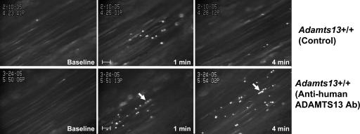
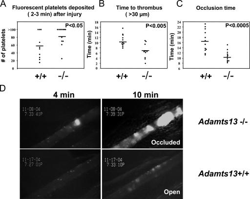
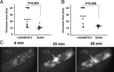
The metalloprotease ADAMTS13 (a disintegrin-like and metalloprotease with thrombospondin type I repeats 13) cleaves highly adhesive large von Willebrand factor (VWF) multimers after their release from the endothelium. ADAMTS13 deficiency is linked to a life-threatening disorder, thrombotic thrombocytopenic purpura (TTP), characterized by platelet-rich thrombi in the microvasculature. Here, we show spontaneous thrombus formation in activated microvenules of Adamts13-/- mice by intravital microscopy. Strikingly, we found that ADAMTS13 down-regulates both platelet adhesion to exposed subendothelium and thrombus formation in injured arterioles. An inhibitory antibody to ADAMTS13 infused in wild-type mice prolonged adhesion of platelets to endothelium and induced thrombi formation with embolization in the activated microvenules. Absence of ADAMTS13 did not promote thrombi formation in alphaIIbbeta3 integrin-inhibited blood. Recombinant ADAMTS13 reduced platelet adhesion and aggregation in histamine-activated venules and promoted thrombus dissolution in injured arterioles. Our findings reveal that ADAMTS13 has a powerful natural antithrombotic activity and recombinant ADAMTS13 could be used as an antithrombotic agent.


Figures

Figure 1.
Figure 1.
Thrombus formation in stimulated microvenules of Adamts13−/− mice. Venules measuring ∼25–30-μm in diameter were visualized in the mesentery of live mice. 1 min after topical superfusion of calcium ionophore A23187, thrombus formation was observed in Adamts13 −/− mice (n = 5). No microthrombi formed in Adamts13 +/+ mice treated identically (n = 5). Arrows indicate the microthrombi. See Video 1 (available at http://www.jem.org/cgi/content/full/jem.20051732/DC1) for thrombi in the microvenules of Adamts13 −/− mice.
Figure 2.
Figure 2.
Antibody to ADAMTS13 increases platelet adhesion and string formation on activated vessel wall. Fluorescently labeled platelets representing ∼2.5% of total platelets were observed in mesenteric venules (diameter: 200–250 μm) of live mice before (baseline) and after A23187 superfusion. Platelets began to adhere to the endothelium 30–45 s after superfusion. In Adamts13 +/+ mice (infused with anti–human ADAMTS13 Ab, n = 4), more platelets adhered to the vessel wall 4 min after stimulation compared with Adamts13 +/+ control (n = 4). Arrows indicate the ≥20-μm strings of platelets attached at one end to the endothelium and waving the other end in the blood stream. Inset time points in the lower right corner refer to the time after superfusion of A23187. The bar shown in the middle panel is ∼50 μm.
Figure 3.
Figure 3.
Thrombus formation in microvenules of Adamts13+/+ mice infused with an anti-ADAMTS13 antibody. Mesenteric venules of ∼25–30-μm in diameter were observed. 1 min after topical superfusion with A23187, thrombus formation was observed in four out of six Adamts13 +/+ mice infused with the anti-ADAMTS13 Ab. The microthrombi formation and embolization were similar to that seen in Adamts13 −/− mice (Fig. 1). Arrows indicate a microthrombus. Microthrombi did not form in Adamts13 +/+ control (n = 5).
Figure 4.
Figure 4.
Recombinant ADAMTS13 inhibits platelet strings in Adamts13−/− mice. Rhodamine 6G was used to label endogenous platelets and leukocytes. Histamine was administered i.p. 15 min before surgery and three mesenteric venules of ∼200–300 μm in diameter were visualized per mouse. (A) No platelet strings are seen in Adamts13 +/+ mice (n = 5). (B) Platelet strings (indicated by arrows) are seen in the Adamts13 −/− mice. Platelet strings anchor up to 1 min on the endothelium (n = 5). (C) The platelet strings could form platelet aggregates in Adamts13 −/− mice as indicated by arrow. (D) Infusion of r-hu ADAMTS13 protein inhibits the platelet strings in Adamts13 −/− mice (n = 4).
Figure 5.
Figure 5.
Quantitative analysis of platelet adhesion and thrombi formation in FeCl3-injured arterioles of Adamts13+/+ and Adamts13−/− mice. (A) The number of fluorescent platelets deposited per minute was determined in the interval 2–3 min after injury. Absence of ADAMTS13 in the plasma significantly increases early platelet interaction with the subendothelium (P < 0.05). (B) Thrombi (>30 μm) appeared sooner in Adamts13 −/− mice compared with Adamts13 +/+ (P < 0.005). (C) The occlusion time (blood flow completely stopped for 10 s) was determined. Both Adamts13 +/+ and Adamts13 −/− mice occluded at the site of injury; however, in Adamts13 −/− mice, occlusion time was shorter as compared with Adamts13 +/+ mice (P < 0.0005). (D) Fluorescently labeled platelets representing ∼2.5% of total platelets were observed in mesenteric arterioles of live mice after FeCl3 injury. Single adherent platelets are seen in the arteriole at 4 min after injury in the Adamts13 +/+ mouse, whereas a thrombus (∼30 μm) can already be seen in the Adamts13 −/− mouse at the same time point. The vessel was occluded at 10 min at the site of injury in the Adamts13 −/− mouse, whereas the Adamts13 +/+ mouse arteriole remained opened at that time. Representative figures are shown. Blood flow was from left to right.
Figure 6.
Figure 6.
Inhibition of integrin αIIbβ3 blocks thrombus formation of ADAMTS13−/− platelets on collagen under arterial shear rate conditions. Adamts13 +/+ or Adamts13 −/− whole blood was perfused for 2 min over a collagen surface at a shear rate of 1, 500 s−1. (A) Representative images are shown. (top) Untreated whole blood; (bottom) whole blood pretreated with blocking antibody against αIIbβ3 (JON/A). (B) Quantification of the surface area covered by platelets after 2 min of perfusion. Four frames from different areas of the flow chamber were analyzed for each blood sample. Data represent the mean percentage of surface area covered by fluorescent platelets ± SEM (n = 3–4).
Figure 7.
Figure 7.
Infusion of r-hu ADAMTS13 inhibits thrombus growth. r-hu ADAMTS13 was infused i.v. into the Adamts13 −/− mice 15 min before the FeCl3 injury. The occlusion time (blood flow completely stopped for 10 s) was determined. (A) 5 out of 13 Adamts13 −/− mice infused with r-hu ADAMTS13 did not occlude in the arteriole at up to 40 min of observation time (mean occlusion time = 23.80 ± 3.71 min), whereas all 10 Adamts13 −/− mice infused with recombinant buffer occluded (mean occlusion time = 11.17 ± 0.87 min, P = 0.005). (B) Occlusion time in injured arterioles of WT (C57BL/6J) mice infused either with r-hu ADAMTS13 (mean occlusion time = 27.99 ± 4.72 min) or buffer (mean occlusion time = 13.12 ± 0.55 min) alone. (C) Representative fluorescent images of an injured arteriole of an Adamts13 −/− mouse treated with r-hu ADAMTS13 are shown. Arrows indicate a dissolving thrombus. See Video 2 (available at http://www.jem.org/cgi/content/full/jem.20051732/DC1) for the effect of r-hu ADAMTS13 on thrombus growth.

References

    1. Sadler, J.E., J.L. Moake, T. Miyata, and J.N. George. 2004. Recent advances in thrombotic thrombocytopenic purpura. Hematology (Am. Soc. Hematol. Educ. Program). 2004:407–423. - PubMed
    1. Moake, J.L. 2002. Thrombotic microangiopathies. N. Engl. J. Med. 347:589–600. - PubMed
    1. Moake, J.L., C.K. Rudy, J.H. Troll, M.J. Weinstein, N.M. Colannino, J. Azocar, R.H. Seder, S.L. Hong, and D. Deykin. 1982. Unusually large plasma factor VIII:von Willebrand factor multimers in chronic relapsing thrombotic thrombocytopenic purpura. N. Engl. J. Med. 307:1432–1435. - PubMed
    1. Furlan, M., R. Robles, M. Galbusera, G. Remuzzi, P.A. Kyrle, B. Brenner, M. Krause, I. Scharrer, V. Aumann, U. Mittler, et al. 1998. von Willebrand factor-cleaving protease in thrombotic thrombocytopenic purpura and the hemolytic-uremic syndrome. N. Engl. J. Med. 339:1578–1584. - PubMed
    1. Furlan, M., R. Robles, M. Solenthaler, M. Wassmer, P. Sandoz, and B. Lammle. 1997. Deficient activity of von Willebrand factor-cleaving protease in chronic relapsing thrombotic thrombocytopenic purpura. Blood. 89:3097–3103. - PubMed

Publication types

MeSH terms