. 2006 Mar 13;172(6):909-21.
doi: 10.1083/jcb.200508130.

Osteoblast differentiation and skeletal development are regulated by Mdm2-p53 signaling

Affiliations

Osteoblast differentiation and skeletal development are regulated by Mdm2-p53 signaling

Christopher J Lengner et al. J Cell Biol. .

Abstract

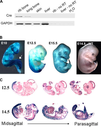
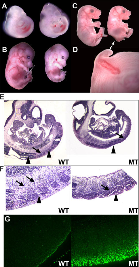
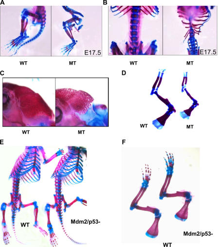
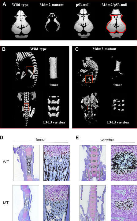
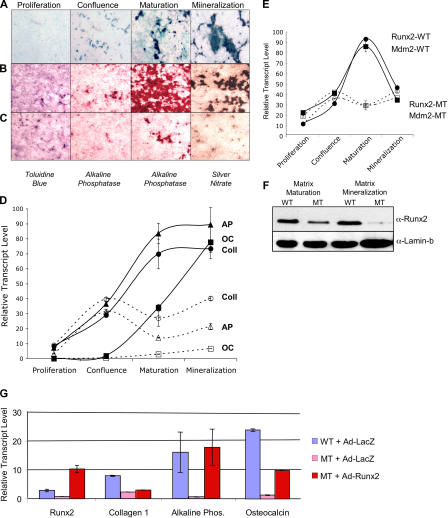
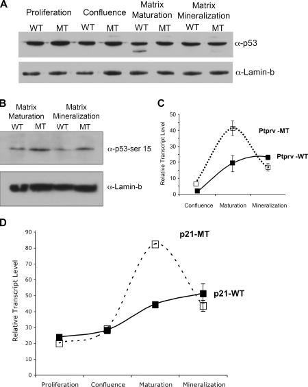
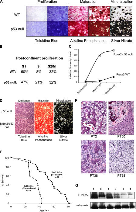
Mdm2 is required to negatively regulate p53 activity at the peri-implantation stage of early mouse development. However, the absolute requirement for Mdm2 throughout embryogenesis and in organogenesis is unknown. To explore Mdm2-p53 signaling in osteogenesis, Mdm2-conditional mice were bred with Col3.6-Cre-transgenic mice that express Cre recombinase in osteoblast lineage cells. Mdm2-conditional Col3.6-Cre mice die at birth and display multiple skeletal defects. Osteoblast progenitor cells deleted for Mdm2 have elevated p53 activity, reduced proliferation, reduced levels of the master osteoblast transcriptional regulator Runx2, and reduced differentiation. In contrast, p53-null osteoprogenitor cells have increased proliferation, increased expression of Runx2, increased osteoblast maturation, and increased tumorigenic potential, as mice specifically deleted for p53 in osteoblasts develop osteosarcomas. These results demonstrate that p53 plays a critical role in bone organogenesis and homeostasis by negatively regulating bone development and growth and by suppressing bone neoplasia and that Mdm2-mediated inhibition of p53 function is a prerequisite for Runx2 activation, osteoblast differentiation, and proper skeletal formation.


Figures

Figure 1.
Figure 1.
Expression of Cre recombinase in Col3.6-Cre–transgenic mice. (A) PCR performed using Cre primers on reverse-transcribed RNA isolated from adult Col3.6-Cre–transgenic mice. Primers to GAPDH were used as a positive control in the RT-PCR, whereas non–reverse-transcribed (No RT) rib bone RNA, liver RNA, and water (H20) were used as negative controls for the PCR. Cre expression is readily detected in the rib and femur of these mice. (B) Whole-mount staining of lacZ expression in Col3.6-Cre–transgenic R26R embryos at E10, -12.5, and -15.5 during development. Expression of the Cre recombinase as detected by β-galactosidase activity is detected in surface ectoderm and in the caudal portion of the embryo at E10, with the highest levels found in the tail bud (arrow). Between E12.5 and -15.5, the Col3.6-Cre transgene undergoes robust activation with the formation of connective tissue. (C) Staining for β-galactosidase activity in sagital sections of R26R/Col3.6-Cre embryos indicates that Cre-mediated excision occurs in both the skin and developing skeletal elements at E12.5–14.5.
Figure 2.
Figure 2.
Caudal defects in Mdm2-conditional Col3.6-Cre mice. (A–D) Homozygous Mdm2-conditional embryos lacking (left) or containing (right) the Col3.6-Cre transgene were harvested from timed matings at E10 (A), E15 (B), and E17.5 (C). Embryos mutated for Mdm2 were generally smaller and displayed obvious runting of the caudal portion of the embryo, absence of a tail, and a severe invagination in the posterior dorsal region encompassing the lumbar vertebrae (D, double-headed arrow). (E and F) Hematoxylin and eosin staining of sagital sections of wild-type (WT) and Mdm2-conditional Col3.6-Cre (MT) embryos (E). Arrows identify somites that are surrounded by primitive neural tissue and by developing dorsal root ganglia. Mutant embryos lack developing neural tissue and posterior dorsal root ganglia (arrowheads), leaving caudal somites externalized (F). (G) TUNEL staining performed on serial sections shown in F demonstrates increased TUNEL-positive apoptotic cells in the caudal somites and surrounding tissue of mutant embryos in comparison to wild-type littermates.
Figure 3.
Figure 3.
Excision of Mdm2 in skeletal tissues results in impaired bone formation. Alcian blue and alizarin red staining of skeletal preparations of wild-type (WT) and Mdm2sjcnd1/sjcnd1, Col3.6-Cre (MT) embryos. (A and B) Mutant embryos harvested at E17.5 revealed highly dysplastic axial skeletal elements with fused cartilaginous lumbar vertebrae. (C) The skulls of Mdm2sjcnd1/sjcnd1,Col3.6-Cre E17.5 embryos are highly porous, and (D) appendicular bones of the forelimb were shorter in length in comparison to wild-type littermates. Skeletal preparations of wild-type neonatal mice and neonates deleted for both Mdm2 and p53 revealed no vertebral dysplasia (E) or reduction in bone length (F), indicating that the deleterious effects of Mdm2 loss on skeletal formation during development are p53 dependent.
Figure 4.
Figure 4.
Altered mineralization and morphology observed in Mdm2 mutant mice. (A) Micro-CT analysis of skulls of wild-type; Mdm2sjcnd1/sjcnd1,Col3.6-Cre (Mdm2 mutant); p53-null; and Mdm2/p53 double-null E18.5 embryos reveals reduced bone mineralization in Mdm2 mutant mice. Histomorphometric measurements of bone volume versus total volume are provided in Table II for each skull, with a representative area analyzed for the Mdm2/p53-null sample outlined in red. Micro-CT scan of femur and vertebra (L3–5 region) of Mdm2 of wild-type E18.5 embryo (B) and Mdm2 mutant embryo (C). Regions of bones analyzed in Table II are outlined in red and shown in expanded view. Von Kossa (silver nitrate) and toluidine blue staining of sagital sections of femur (D) from Mdm2-conditional Col3.6-Cre (MT) mice showed a relatively normal growth plate region but had significantly less mineral deposition than Mdm2-conditional mice lacking the Cre transgene (WT). Similarly, less mineralization was also detected in Von Kossa and toluidine blue staining (E) of Mdm2-conditional Col3.6-Cre vertebra compared with wild-type vertebra.
Figure 5.
Figure 5.
Mdm2 is required for proper osteoblast differentiation. Calvarial osteoprogenitor cells were isolated and cultured ex vivo from E19 Col3.6-Cre–transgenic embryos bearing the R26R reporter gene, from E19 wild-type and Mdm2-conditional Col3.6-Cre–transgenic embryos. Calvarial osteoprogenitors were induced to undergo osteogenic differentiation ex vivo, resulting in proliferation (toluidine blue staining for total cell number), the formation of multilayered nodules and activation of alkaline phosphatase, and mineralization as detected by silver nitrate staining. (A) Examination R26R/Col3.6-Cre osteoprogenitor cells stained for β-galactosidase activity reveals that Mdm2 expression in Mdm2-conditional Col3.6-Cre cultures will be lost in multilayering nodules of maturing osteoblasts. (B) Upon reaching confluence, wild- type osteoprogenitor cell cultures underwent robust nodule formation and mineralization. (C) Mdm2-conditional Col3.6-Cre–transgenic cultures were unable to form a significant number of multilayered nodules, and subsequently only a small fraction of these cells were able to undergo differentiation, activate alkaline phosphatase activity, or induce mineralization. (D) Quantitative analysis by real-time PCR of the expression of early osteogenic genes type I collagen (Coll) and alkaline phosphatase (AP) and of osteocalcin (OC) at various stages of osteoblast maturation. Solid lines depict the relative transcript levels of genes in wild-type cell cultures, and dashed lines represent the levels of expression of the various genes in Mdm2-conditional Col3.6-Cre cell cultures. Expression levels of early and late osteogenic genes are reduced in Mdm2-conditional Col3.6-Cre–transgenic cells. (E) Real-time PCR analysis of Mdm2 and Runx2 expression in wild-type (WT) and Mdm2-conditional Col3.6-Cre–transgenic (MT) culture osteoblast progenitor cells during maturation. Solid and dashed lines depict the relative transcript levels of Mdm2 and Runx2 in wild-type or Mdm2-conditional Col3.6-Cre–transgenic cultures, respectively. As expected, Mdm2 levels do not increase in Mdm2-conditional Col3.6-Cre–transgenic cells during maturation. Similar induction of Runx2 and Mdm2 expression is observed in wild-type cells during maturation. (F) Western blot analysis of Runx2 protein levels in wild-type or Mdm2-conditional Col3.6-Cre–transgenic culture osteoblast progenitor cells during maturation and mineralization. Lamin-b is shown as a loading control. Decreased amounts of Runx2 protein are observed in Mdm2-conditional Col3.6-Cre–transgenic cells. (G) Quantitative PCR was performed on reverse-transcribed RNA isolated from osteoblast progenitor cells transduced with Runx2 or lacZ (negative control). Exogenous Runx2 up-regulated the expression of osteogenic maturation genes in Mdm2 mutant cells, including collagen 1, alkaline phosphatase, and osteocalcin. Error bars indicate SD.
Figure 6.
Figure 6.
Deletion of Mdm2 alters p53 activity in osteoblast cultures. (A) Western analysis of p53 levels in wild-type (WT) and Mdm2-conditional Col3.6-Cre–transgenic (MT) osteoprogenitor cells during differentiation reveals no change in total p53 protein levels in cultures undergoing deletion of Mdm2. (B) Western analysis of phosphorylated p53 in wild-type and Mdm2-conditional Col3.6-Cre–transgenic cells during maturation and mineralization reveals increased amounts of activated p53 in MT cells. Lamin-b is shown as a loading control. (C and D) Real-time PCR analysis of RNA isolated from wild-type or Mdm2-conditional Col3.6-Cre–transgenic cells during differentiation reveals up-regulation of Pptrv or p21 gene expression in postconfluent osteoblast cultures derived from Mdm2-conditional Col3.6-Cre mice (dashed line) relative to expression levels in cells containing wild-type Mdm2 alleles (solid line). Error bars indicate SD.
Figure 7.
Figure 7.
Regulation of osteoblast progenitor differentiation and osteosarcoma formation by p53. Calvarial osteoprogenitor cells were cultured ex vivo from E19 wild-type (WT) and p53-null embryos. (A) Calvarial osteoprogenitors were induced to undergo osteogenic differentiation, resulting in proliferation (toluidine blue), maturation and the formation multilayered nodules and activation of alkaline phosphatase, and mineralization as detected by silver nitrate staining. Osteoprogenitor cells deleted for p53 displayed far more proliferation than wild-type cells cultured at the same initial density, increased alkaline phosphatase, and increased mineralization. (B) Proliferation was documented by BrdU uptake in postconfluent cultures of wild-type and p53-null progenitor cells just before cell maturation. The percentage of cells in each phase of the cell cycle was determined by FACS analysis after propidium iodide staining of the harvested cells. Postconfluent p53-null cells had more cells in S phase than wild-type cells. (C) Real-time PCR analysis of Runx2 expression in wild-type and p53-null cultures of osteoblast progenitor cells during maturation. Runx2 transcript levels are strongly up-regulated in p53-null cells during maturation. (D) No difference was observed in the robust osteoprogenitor cell differentiation in p53-null cells that contained (top) or lacked (bottom) Mdm2. (E) Tumorigenesis in mice deleted for p53 in osteoblasts. Col3.6-Cre–transgenic mice heterozygous for a conditional p53 allele (p53-cond/wild type) or homozygous for the p53-conditional allele (p53-cond/cond) were assayed for spontaneous tumor development. A majority of the mice presented with osteosarcomas, with a mean time to tumorigenesis of 40 wk for Col3.6-Cre, p53-cond/cond mice and 57 wk for Col3.6-Cre, p53-cond/wild-type mice. (F) Hematoxylin and eosin stains of osteosarcomas harvested from Col3.6-Cre, p53-cond/cond mice. Samples PT2, -50 (showing invasion into the liver), and -38 are all more differentiated than sample PT58, which displays a spindle-like morphology in addition to some osteoid cells. (G) Analysis of Runx2 protein levels in representative primary tumor samples from p53-conditional Col3.6-Cre mice. Runx2 levels were readily detected in representative osteosarcomas (lanes 1–3: samples PT2, -38, and -58, respectively) but reduced in fibrosarcoma (lane 4), lymphoma (lane 5), or hemangiosarcoma (lane 6) samples.

Comment in

References

    1. Alt, J.R., T.C. Greiner, J.L. Cleveland, and C.M. Eischen. 2003. Mdm2 haplo-insufficiency profoundly inhibits Myc-induced lymphomagenesis. EMBO J. 22:1442–1450. - PMC - PubMed
    1. Armstrong, J.F., M.H. Kaufman, D.J. Harrison, and A.R. Clarke. 1995. High-frequency developmental abnormalities in p53-deficient mice. Curr. Biol. 5:931–936. - PubMed
    1. Banerjee, C., L.R. McCabe, J.-Y. Choi, S.W. Hiebert, J.L. Stein, G.S. Stein, and J.B. Lian. 1997. Runt homology domain proteins in osteoblast differentiation: AML3/CBFA1 is a major component of a bone-specific complex. J. Cell. Biochem. 66:1–8. - PubMed
    1. Chen, J., J. Lin, and A.J. Levine. 1995. Regulation of transcription functions of the p53 tumor suppressor by the mdm-2 oncogene. Mol. Med. 1:142–152. - PMC - PubMed
    1. Deng, C., P. Zhang, J.W. Harper, S.J. Elledge, and P. Leder. 1995. Mice lacking p21CIP1/WAF1 undergo normal development, but are defective in G1 checkpoint control. Cell. 82:675–684. - PubMed

Publication types

MeSH terms

Substances