

Memory fMRI in left hippocampal sclerosis: optimizing the approach to predicting postsurgical memory
- PMID: 16534106
- PMCID: PMC2649428
- DOI: 10.1212/01.wnl.0000201186.07716.98
Memory fMRI in left hippocampal sclerosis: optimizing the approach to predicting postsurgical memory
Abstract
Background: An optimal technique for clinical memory fMRI is not established. Previous studies suggest activity in right parahippocampal gyrus and right hippocampus shows the strongest difference between left hippocampal sclerosis (HS) patients and normal control subjects and that the difference in activity between left and right hippocampus predicts postoperative memory change.
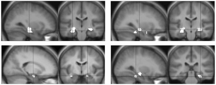
Methods: The authors studied 30 patients with mesial temporal lobe epilepsy (mTLE) and left HS, 12 of whom subsequently underwent surgery, and 13 normal control subjects. The patients who had surgery underwent neuropsychometric evaluation pre- and postoperatively. All subjects underwent a verbal memory encoding event-related fMRI study. Activation maps were assessed visually. Subsequently, the brain regions involved in the memory task were revealed by group averaging; these regions were used to determine regions of interest (ROIs) for subsequent analysis. By use of stepwise discriminant function and stepwise multiple regression, the ROIs that optimally discriminated between patients and normal control subjects and that optimally predicted postoperative verbal memory outcome were determined.
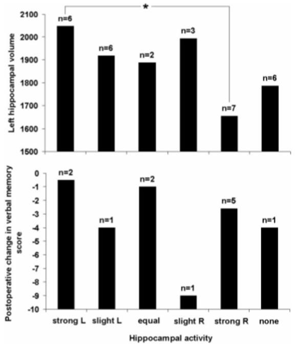
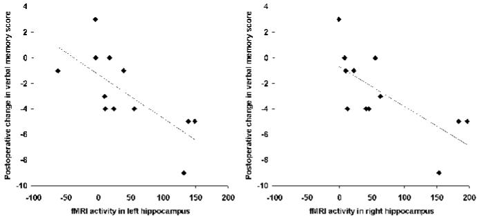
Results: Visual inspection of individual patient activation statistic maps revealed noisy data that did not afford visual interpretation. Stepwise discriminant function revealed the difference between left and right hippocampal activity best discriminated between patients and normal control subjects. Stepwise multiple regression revealed left hippocampal activity was the strongest predictor of postoperative verbal memory outcome; greater left hippocampal activity predicted a greater postoperative decline in memory.
Conclusions: Patients with left hippocampal sclerosis (HS) differ from normal control subjects in the distribution of memory-encoding activity between left and right hippocampus. Functional adequacy of left hippocampus best predicts postoperative memory outcome in left HS.
Figures
Comment in
-
How to image memory in epilepsy.Epilepsy Curr. 2006 Nov-Dec;6(6):189-91. doi: 10.1111/j.1535-7511.2006.00141.x. Epilepsy Curr. 2006. PMID: 17260055 Free PMC article. No abstract available.
References
-
- Plate KH, Wieser HG, Yasargil MG, Wiestler OD. Neuropathological findings in 224 patients with temporal lobe epilepsy. Acta Neuropathol Berl. 1993;86:433–438. - PubMed
-
- Kuzniecky R, Murro A, King D, et al. Magnetic resonance imaging in childhood intractable partial epilepsies: pathologic correlations. Neurology. 1993;43:681–687. - PubMed
-
- Wolf HK, Campos MG, Zentner J, et al. Surgical pathology of temporal lobe epilepsy. Experience with 216 cases. J Neuropathol Exp Neurol. 1993;52:499–506. - PubMed
-
- Wiebe S, Blume WT, Girvin JP, Eliasziw M. A randomized, controlled trial of surgery for temporal-lobe epilepsy. N Engl J Med. 2001;345:311–318. - PubMed
-
- Katz A, Awad IA, Kong AK, et al. Extent of resection in temporal lobectomy for epilepsy. II. Memory changes and neurologic complications. Epilepsia. 1989;30:763–771. - PubMed
Publication types
MeSH terms
Grants and funding
LinkOut - more resources
Full Text Sources
Medical