Primary liposarcoma of esophagus: a case report
- PMID: 16534863
- PMCID: PMC4087914
- DOI: 10.3748/wjg.v12.i7.1149
Primary liposarcoma of esophagus: a case report
Abstract
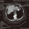
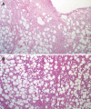
Liposarcoma is the most common soft tissue sarcoma in adult life while esophageal liposarcoma is an extremely rare tumor. In the world literature, only 14 cases of esophageal liposarcomas have been described. We report a 72-year old male patient who was urgently admitted to our hospital for acute epigastric pain with a burning retrosternal sensation, persistent nausea, vomiting and dysphagia. Barium swallow, upper gastrointestinal (GI) endoscopy, esophageal manometry and CT scan, failed to accurately diagnose the lesion. After surgical resection of an esophageal polypoid tumor, the histological examination revealed a well-differentiated grade I liposarcoma. Diagnostic and therapeutic tools were discussed and the results of literature were reviewed.
Figures


References
-
- Weiss SW, Goldblum JR. Liposarcoma, in Weiss SW, Goldblum JR. eds. Enzinger and Weiss's. 4th ed. Philadelphia: Mosby Harcourt; 2001. pp. 641–662.
-
- Mansour KA, Fritz RC, Jacobs DM, Vellios F. Pedunculated liposarcoma of the esophagus: a first case report. J Thorac Cardiovasc Surg. 1983;86:447–450. - PubMed
-
- Garcia M, Buitrago E, Bejarano PA, Casillas J. Large esophageal liposarcoma: a case report and review of the literature. Arch Pathol Lab Med. 2004;128:922–925. - PubMed
-
- Yates SP, Collins MC. Case report: recurrent liposarcoma of the oesophagus. Clin Radiol. 1990;42:356–358. - PubMed
Publication types
MeSH terms
LinkOut - more resources
Full Text Sources
Medical
Molecular Biology Databases

