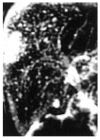
Bile duct hamartomas (von Mayenburg complexes) mimicking liver metastases from bile duct cancer: MRC findings
- PMID: 16534895
- PMCID: PMC4124453
- DOI: 10.3748/wjg.v12.i8.1321
Bile duct hamartomas (von Mayenburg complexes) mimicking liver metastases from bile duct cancer: MRC findings
Abstract
We present a case of a 72-year-old man with a common bile duct cancer, who was initially believed to have multiple liver metastases based on computed tomography findings, and in whom magnetic resonance cholangiography (MRC) revealed a diagnosis of bile duct hamartomas. At exploration for pancreaticoduodenectomy, liver palpation revealed disseminated nodules at the surface of the liver. These nodules showed gray-white nodular lesions of about 0.5 cm in diameter scattered on the surface of both liver lobes, which were looked like multiple liver metastases from bile duct cancer. Frozen section of the liver biopsy disclosed multiple bile ducts with slightly dilated lumens embedded in the collagenous stroma characteristics of multiple bile duct hamartomas (BDHs). Only two reports have described the MRC features of bile duct hamartomas. Of all imaging procedures, MRC provides the most relevant features for the imaging diagnosis of bile duct hamartomas.
Figures


References
-
- Wei SC, Huang GT, Chen CH, Sheu JC, Tsang YM, Hsu HC, Chen DS. Bile duct hamartomas. A report of two cases. J Clin Gastroenterol. 1997;25:608–611. - PubMed
-
- Eisenberg D, Hurwitz L, Yu AC. CT and sonography of multiple bile-duct hamartomas simulating malignant liver disease (case report) AJR Am J Roentgenol. 1986;147:279–280. - PubMed
-
- Luo TY, Itai Y, Eguchi N, Kurosaki Y, Onaya H, Ahmadi Y, Niitsu M, Tsunoda HS. Von Meyenburg complexes of the liver: imaging findings. J Comput Assist Tomogr. 1998;22:372–378. - PubMed
-
- Mortelé B, Mortelé K, Seynaeve P, Vandevelde D, Kunnen M, Ros PR. Hepatic bile duct hamartomas (von Meyenburg Complexes): MR and MR cholangiography findings. J Comput Assist Tomogr. 2002;26:438–443. - PubMed
-
- Chamlou R, Matos C, Nagy N, Gelin M, Closset J. Pitfalls on frozen section of a hepatic lesion in the management of a pancreatic tumor. Surgery. 2003;133:599–600. - PubMed
Publication types
MeSH terms
LinkOut - more resources
Full Text Sources
Medical

