

Role of MAP1B in axonal retrograde transport of mitochondria
- PMID: 16536727
- PMCID: PMC1479764
- DOI: 10.1042/BJ20060205
Role of MAP1B in axonal retrograde transport of mitochondria
Abstract
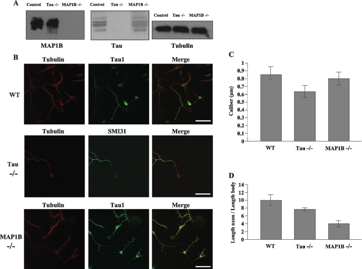
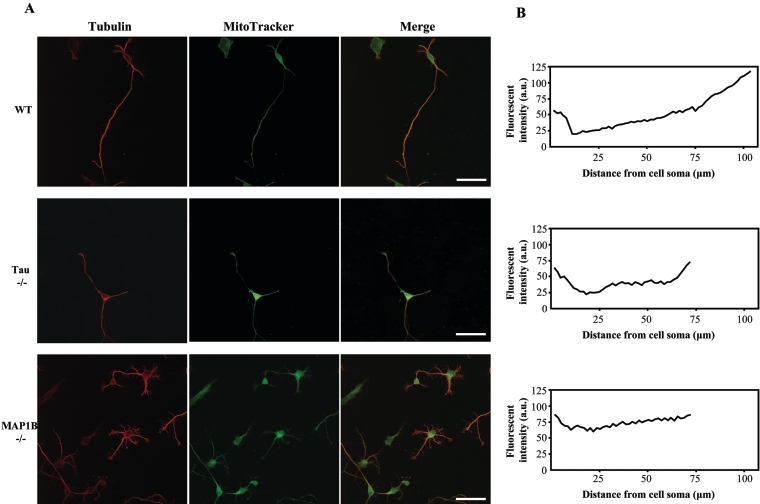
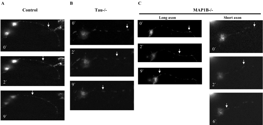
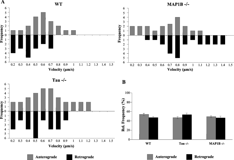
The MAPs (microtubule-associated proteins) MAP1B and tau are well known for binding to microtubules and stabilizing these structures. An additional role for MAPs has emerged recently where they appear to participate in the regulation of transport of cargos on the microtubules found in axons. In this role, tau has been associated with the regulation of anterograde axonal transport. We now report that MAP1B is associated with the regulation of retrograde axonal transport of mitochondria. This finding potentially provides precise control of axonal transport by MAPs at several levels: controlling the anterograde or retrograde direction of transport depending on the type of MAP involved, controlling the speed of transport and controlling the stability of the microtubule tracks upon which transport occurs.
Figures
Similar articles
-
Synergistic effects of MAP2 and MAP1B knockout in neuronal migration, dendritic outgrowth, and microtubule organization.J Cell Biol. 2001 Oct 1;155(1):65-76. doi: 10.1083/jcb.200106025. J Cell Biol. 2001. PMID: 11581286 Free PMC article.
-
Acute inactivation of MAP1b in growing sympathetic neurons destabilizes axonal microtubules.Cell Motil Cytoskeleton. 2005 Jan;60(1):48-65. doi: 10.1002/cm.20045. Cell Motil Cytoskeleton. 2005. PMID: 15573412
-
A possible mechanism for controlling processive transport by microtubule-associated proteins.Neurosci Res. 2008 Aug;61(4):347-50. doi: 10.1016/j.neures.2008.04.010. Epub 2008 May 2. Neurosci Res. 2008. PMID: 18541318 Review.
-
Defects in axonal elongation and neuronal migration in mice with disrupted tau and map1b genes.J Cell Biol. 2000 Sep 4;150(5):989-1000. doi: 10.1083/jcb.150.5.989. J Cell Biol. 2000. PMID: 10973990 Free PMC article.
-
Neuronal microtubules: when the MAP is the roadblock.Trends Cell Biol. 2005 Apr;15(4):183-7. doi: 10.1016/j.tcb.2005.02.001. Trends Cell Biol. 2005. PMID: 15817373 Review.
Cited by
-
Fractalkine signaling and Tau hyper-phosphorylation are associated with autophagic alterations in lentiviral Tau and Aβ1-42 gene transfer models.Exp Neurol. 2014 Jan;251:127-38. doi: 10.1016/j.expneurol.2013.01.009. Epub 2013 Jan 16. Exp Neurol. 2014. PMID: 23333589 Free PMC article.
-
The neurogenic basic helix-loop-helix transcription factor NeuroD6 concomitantly increases mitochondrial mass and regulates cytoskeletal organization in the early stages of neuronal differentiation.ASN Neuro. 2009 Sep 16;1(4):e00016. doi: 10.1042/AN20090036. ASN Neuro. 2009. PMID: 19743964 Free PMC article.
-
MAP1B-LC1 prevents autophagosome formation by linking syntaxin 17 to microtubules.EMBO Rep. 2018 Aug;19(8):e45584. doi: 10.15252/embr.201745584. Epub 2018 Jun 19. EMBO Rep. 2018. PMID: 29925525 Free PMC article.
-
GSK-3β, a pivotal kinase in Alzheimer disease.Front Mol Neurosci. 2014 May 21;7:46. doi: 10.3389/fnmol.2014.00046. eCollection 2014. Front Mol Neurosci. 2014. PMID: 24904272 Free PMC article. Review.
-
MAP1B Light Chain Modulates Synaptic Transmission via AMPA Receptor Intracellular Trapping.J Neurosci. 2017 Oct 11;37(41):9945-9963. doi: 10.1523/JNEUROSCI.0505-17.2017. Epub 2017 Sep 13. J Neurosci. 2017. PMID: 28904092 Free PMC article.
References
-
- Matus A. Microtubule-associated proteins: their potential role in determining neuronal morphology. Annu. Rev. Neurosci. 1988;11:29–44. - PubMed
-
- Mitchison T., Kirschner M. Cytoskeletal dynamics and nerve growth. Neuron. 1988;1:761–772. - PubMed
-
- Avila J., Dominguez J., Díaz-Nido J. Regulation of microtubule dynamics by microtubule-associated protein expression and phosphorylation during neuronal development. Int. J. Dev. Biol. 1994;38:13–25. - PubMed
-
- Tucker R. P., Garner C. C., Matus A. In situ localization of microtubule-associated protein mRNA in the developing and adult rat brain. Neuron. 1989;2:1245–1256. - PubMed
-
- Binder L. I., Frankfurter A., Rebhun L. I. Differential localization of MAP-2 and tau in mammalian neurons in situ. Ann. N.Y. Acad. Sci. 1986;466:145–166. - PubMed
Publication types
MeSH terms
Substances
LinkOut - more resources
Full Text Sources
Molecular Biology Databases