. 2006 Jun 15;396(3):461-8.
doi: 10.1042/BJ20051690.

A direct interaction with calponin inhibits the actin-nucleating activity of gelsolin

Affiliations

A direct interaction with calponin inhibits the actin-nucleating activity of gelsolin

Imen Ferjani et al. Biochem J. .

Abstract

Gelsolin and calponin are well-characterized cytoskeletal proteins that are abundant and widely expressed in vertebrate tissues. It is also becoming apparent, however, that they are involved in cell signalling. In the present study, we show that gelsolin and calponin interact directly to form a high-affinity (K(d)=16 nM) 1:1 complex, by the use of fluorescent probes attached to both proteins, by affinity chromatography and by immunoprecipitation. These methods show that gelsolin can form high-affinity complexes with two calponin isoforms (basic h1 and acidic h3). They also show that gelsolin binds calponin through regions that have been identified previously as being calponin's actin-binding sites. Moreover, gelsolin does not interact with calponin while calponin is bound to F-actin. Reciprocal experiments to find calponin-binding sites on gelsolin show that these are in both the N- and C-terminal halves of gelsolin. Calponin has minimal effects on actin severing by gelsolin. In contrast, calponin markedly affects the nucleation activity of gelsolin. The maximum inhibition of nucleation by gelsolin was 50%, which was achieved with a ratio of two calponins for every gelsolin. Thus the interaction of calponin with gelsolin may play a regulatory role in the formation of actin filaments through modulation of gelsolin's actin-binding function and through the prevention of calponin's actin-binding activities.


Figures
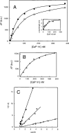
Figure 1
Figure 1. Binding of Oregon-Green-labelled gelsolin to calponin h1
(A) Interaction of Oregon-Green-labelled gelsolin (85 nM) with calponin (CaP) h1 was monitored by fluorescence. Changes in the intensity of the fluorescence emission spectra (ΔF) of Oregon Green were recorded at various calponin concentrations (0–380 nM) in 0.1 M Tris/HCl buffer, pH 7.5, supplemented with 1 mM EGTA (●) or 1 mM Ca2+ (○). Inset, blue shift in the maximum of the emission spectra was monitored with respect to calponin concentration (0.1 M Tris/HCl buffer, pH 7.5, containing 1 mM EGTA). a.u., arbitrary units. (B) Interaction of Oregon-Green-labelled gelsolin (85 nM) with calponin (CaP) h1 in 0.1 M Mes buffer, pH 6.0. Calponin h1 concentration was varied between 0 and 480 nM. a.u., arbitrary units. (C) Quantitative analysis of the data in (A) and (B) for the interaction between gelsolin and calponin h1 was performed by plotting 1/(1−X) against C/X·E where C is the concentration of calponin expressed in nM and E is the concentration of gelsolin fixed at 85 nM. X, the binding ratio, was determined as described in the Materials and methods section. The experiments were performed in 0.1 M Tris/HCl buffer, pH 7.5, supplemented with EGTA (●) or Ca2+ (○), or in 0.1 M Mes buffer, pH 6.0 (□).
Figure 2
Figure 2. Interaction of acrylodan-labelled calponin h1 with gelsolin
Interaction of gelsolin with calponin was monitored by changes in acrylodan fluorescence intensity. Experiments were carried out in 0.1 M Tris/HCl buffer, pH 7.5, and 1 mM EGTA using a fixed concentration of labelled calponin of 80 nM. a.u., arbitrary units. (A) Emission spectrum of acrylodan-labelled calponin alone (——) or in the presence of 48 nM gelsolin (− − −). The excitation wavelength was 388 nm. (B) Fluorescence decrease is plotted against gelsolin concentrations varied between 0 and 200 nM. (C) Quantitative analysis of the data in (B) for the interaction between acrylodan-labelled calponin h1 and gelsolin was performed by plotting 1/(1−X) against C/X·E, where C is the concentration of gelsolin (in nM) and E is the concentration of calponin h1 fixed at 80 nM.
Figure 3
Figure 3. Binding of gelsolin to calponin h1 coupled to Sepharose 4B
Gelsolin (870 μg) was passed through a column (1.4 cm×3 cm) of Sepharose-4B-linked calponin. The column was washed with 10 mM phosphate buffer, pH 7.4, supplemented with 0.15 M NaCl. The bound material was eluted with 10 mM phosphate buffer, pH 12, supplemented with 10% dioxan. The eluted fraction was quantified from a UV absorption spectrum (in a typical experiment, 780 μg of gelsolin was eluted), pulled-down and analysed by SDS/12.5% PAGE (inset). Lane 1, gelsolin control; lane 2, eluted material.
Figure 4
Figure 4. Gelsolin and calponin interact in vivo
(A) Co-localization of gelsolin and calponin in COS-7 cells. Endogenous calponin was revealed by an anti-(calponin h3) antibody followed by an FITC-conjugated anti-rabbit antibody (a); endogenous gelsolin was revealed by an anti-gelsolin antibody followed by a TRITC-conjugated anti-mouse antibody (b); co-localization of the two proteins (in yellow) is presented in (c). (B) Co-immunoprecipitation of gelsolin and calponin. RIPA extracts were subjected to immunoprecipitation with an anti-(calponin h3) antibody (lane 1), control (lane 2) and an anti-gelsolin antibody (lane 3) followed by Western blots with the indicated antibodies.
Figure 5
Figure 5. Binding of Oregon-Green-labelled gelsolin domains to calponin h1
Interaction of Oregon-Green-labelled G1–G3 (140 nM) (○) and G4–G6 (89 nM) (●) with calponin (CaP) h1 was monitored by fluorescence. Changes in the intensity of the fluorescence emission spectra (ΔF) of Oregon Green were recorded at various calponin concentrations (0–280 nM) in 0.1 M Tris/HCl buffer, pH 7.5, supplemented with 1 mM EGTA. Inset, quantitative analysis of the data for the interaction between gelsolin domains and calponin h1 was performed by plotting 1/(1−X) against C/X·E where C is the concentration of calponin (in nM) and E is the concentration of gelsolin domains at the fixed concentration indicated above. Experiments were performed with G1–G3 (○) and G4–G6 (●). a.u., arbitrary units.
Figure 6
Figure 6. Binding of calponin to F-actin in the presence of gelsolin
F-actin (8.5 μM) was incubated in the presence of calponin (2.0 μM) and gelsolin (0.22 μM) in 2 mM MgCl2, 0.1 M KCI, 0.1 mM ATP, 1 mM EGTA and 10 mM Tris/HCl, pH 7.5. The samples were ultracentrifuged at 90000 rev./min for 20 min in a Beckman TLA 100.3 rotor, and the supernatant (S) and the pellet (P) were analysed by SDS/12.5% PAGE. Lanes 1, calponin alone; lanes 2, F-actin alone; lanes 3, F-actin+gelsolin; lanes 4, F-actin+calponin; lanes 5, F-actin+calponin+gelsolin. Molecular-mass markers in kDa are rabbit muscle phosphorylase B (97 kDa), BSA (66 kDa), ovalbumin (45 kDa) and bovine carbonate anhydrase (31 kDa).
Figure 7
Figure 7. Binding of calponin sequences 145–163 and 172–187 to gelsolin monitored by fluorescence (A) and ELISA (B)
(A) The interaction of the 145–163 (○) and 172–187 (●) peptide fragments in 0.1 M Tris/HCl buffer, pH 7.5, supplemented with 1 mM EGTA was monitored by following the corresponding changes in the tryptophan fluorescence (ΔF) of gelsolin (0.2 μM). a.u., arbitrary units. (B) Coated peptides of sequence 145–163 (●) and 172–187 (○) were reacted in 0.1 M Tris/HCl buffer, pH 7.5, and 1 mM EGTA with biotinylated gelsolin at the concentrations indicated.
Figure 8
Figure 8. Effects of calponin on the activation of actin-nucleating activity by gelsolin
(A) Labelled G-actin (9.2 μM) was added to 2 mM MgCl2, 0.1 M KCl, 50 μM ATP, 0.5 mM CaCl2 and 5 mM Tris/HCl, pH 7.5, in the presence of gelsolin (0.23 μM) or gelsolin (0.23 μM)+1–10 molar excess of calponin. In each case, the initial polymerization rate was determined and plotted against the calponin (CaP) h1/gelsolin ratio. a.u., arbitrary units. (B) Labelled G-actin (4.9 μM) was added to 2 mM MgCl2, 0.1 M KCl, 50 μM ATP, 0.5 mM CaCl2 and 5 mM Tris/HCl, pH 7.5, in the absence (——) or the presence of 0.12 μM gelsolin (– – –) or 0.12 μM gelsolin+0.26 μM calponin (−·−·−·), as described in the Materials and methods section. The increase in pyrene fluorescence was plotted against time. Results are means for ten experiments. a.u., arbitrary units.
Figure 9
Figure 9. Effects of calponin on the severing activity of gelsolin
Labelled F-actin (36 μM) (pre-capped by a 1:2000 molar ratio of gelsolin) was diluted to 1.4 μM in 0.5 mM CaCl2, 0.1 mM ATP and 5 mM Tris/HCl, pH 7.5, alone (——) and in the presence of 0.1 μM gelsolin (– – –) or 0.1 μM gelsolin+0.2 μM calponin h1 (−·−·−·). The decrease in fluorescence of pyrenyl actin was plotted against time. a.u., arbitrary units.

Similar articles

Cited by

References

    1. dos Remedios C. G., Chhabra D., Kekic M., Dedova I. V., Tsubakihara M., Berry D. A., Noseworthy N. J. Actin binding proteins: regulation of cytoskeletal microfilaments. Physiol. Rev. 2003;83:433–473. - PubMed
    1. Yin H. L., Stossel T. P. Control of cytoplasmic actin gel–sol transformation by gelsolin, a calcium dependent regulatory protein. Nature (London) 1979;281:583–586. - PubMed
    1. Lamb J. A., Allen P. G., Tuan B. Y., Janmey P. A. Modulation of gelsolin function: activation at low pH overrides Ca2+ requirement. J. Biol. Chem. 1993;268:8999–9004. - PubMed
    1. Lagarrigue E., Ternent D., Maciver S. K., Fattoum A., Benyamin Y., Roustan C. The activation of gelsolin by low pH: the calcium latch is sensitive to calcium but not pH. Eur. J. Biochem. 2003;270:4105–4112. - PubMed
    1. Wang Q., Xie Y., Du Q.-S., Wu X.-J., Feng X., Mei L., McDonald J. M., Xiong W.-C. Regulation of the formation of osteoclastic actin rings by proline-rich tyrosine kinase 2 interacting with gelsolin. J. Cell Biol. 2003;160:565–575. - PMC - PubMed

Publication types