. 2006 Mar 21;103(12):4586-91.
doi: 10.1073/pnas.0505379103. Epub 2006 Mar 13.

Molecular basis for the preferential cleft recognition by dromedary heavy-chain antibodies

Affiliations

Molecular basis for the preferential cleft recognition by dromedary heavy-chain antibodies

Erwin De Genst et al. Proc Natl Acad Sci U S A. .

Abstract

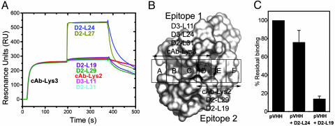
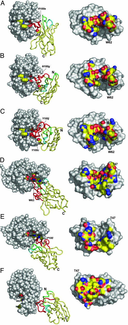
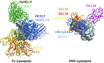
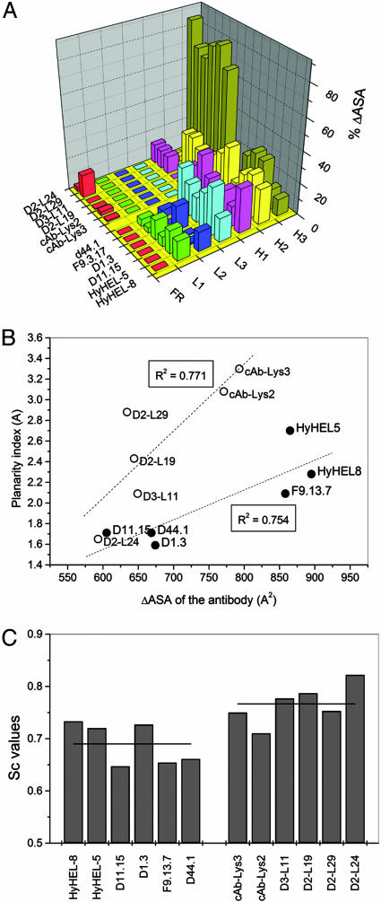
Clefts on protein surfaces are avoided by antigen-combining sites of conventional antibodies, in contrast to heavy-chain antibodies (HCAbs) of camelids that seem to be attracted by enzymes' substrate pockets. The explanation for this pronounced preference of HCAbs was investigated. Eight single domain antigen-binding fragments of HCAbs (VHH) with nanomolar affinities for lysozyme were isolated from three immunized dromedaries. Six of eight VHHs compete with small lysozyme inhibitors. This ratio of active site binders is also found within the VHH pool derived from polyclonal HCAbs purified from the serum of the immunized dromedary. The crystal structures of six VHHs in complex with lysozyme and their interaction surfaces were compared to those of conventional antibodies with the same antigen. The interface sizes of VHH and conventional antibodies to lysozyme are very similar as well as the number and chemical nature of the contacts. The main difference comes from the compact prolate shape of VHH that presents a large convex paratope, predominantly formed by the H3 loop and interacting, although with different structures, into the concave lysozyme substrate-binding pocket. Therefore, a single domain antigen-combining site has a clear structural advantage over a conventional dimeric format for targeting clefts on antigenic surfaces.


Conflict of interest statement

Conflict of interest statement: No conflicts declared.

Figures

Fig. 1.
Fig. 1.
Epitope mapping of the monoclonal and polyclonal VHHs. (A) HEWL binding of all VHHs in the presence of a saturating concentration of cAb-Lys-3 using a coinjection procedure. (B) Epitopes of HEWL active site binders based on the inhibition of binding by NAG3 and Biebrich Scarlet. (C) Residual HEWL binding of polyclonal VHH derived from the IgG3 fraction of dromedary D2. (Left) Binding in absence of competitor (set at 100%). (Center) Binding in presence of HEWL-saturating concentrations of D2-L24. (Right) Binding in presence of HEWL-saturating concentrations of D2-L19.
Fig. 2.
Fig. 2.
The VHH::HEWL complex structures. The D2-L29::HEWL (A), D2-L19::HEWL (B), cAb-Lys-2::HEWL (C), cAb-Lys-3::HEWL (D), D3-L11::HEWL (E), and D2-L24::HEWL (F) complexes are shown. (Left) VHH::HEWL complexes are represented with HEWL molecules as gray space-filling models. The active site residues E35, D52, W62, and W63 are labeled and side-chain atoms color-coded: C, yellow; N, blue; and O, red, for all panels. For AC, the residue T47 of HEWL is additionally colored and labeled. The VHHs are represented as ribbons, with the framework region painted in yellow and the H1, H2, and H3 loops painted in green, cyan, and red respectively. The most important amino acids that contact E35, D52, W62, or W63 of HEWL are represented as sticks and are labeled (italic). (Right) The surface on HEWL within 5-Å distance of the VHH is color-coded according to the atom type: C, yellow; N, blue; O, red; S, orange.
Fig. 3.
Fig. 3.
Superposition of the antibody–lysozyme complexes for conventional antibodies (Left) and for VHHs (Right). The HEWL molecules (gray surfaces) are shown in the same orientation for both antibody classes. The antibody molecules are represented as colored ribbons and their identity is indicated in the same color.
Fig. 4.
Fig. 4.
Structural and chemical differences between VHH::HEWL and Fv::lysozyme interfaces. (A) Contribution of each antigen-binding loop to the ΔASA of the paratope. (B) The planarity index of the VHH or Fv paratope versus their ΔASA. The dotted lines represent the linear trends for both types of antibodies (VHH or Fv) and the Pearson correlation coefficient is indicated. (C) The Sc values for all VHH::HEWL and Fv::lysozyme complexes. The horizontal line through the bars represents the average Sc value for Fvs and VHHs.

References

    1. Padlan E. A. Adv. Protein Chem. 1996;49:57–133. - PubMed
    1. Jones S., Thornton J. M. Proc. Natl. Acad. Sci. USA. 1996;93:13–20. - PMC - PubMed
    1. MacCallum R. M., Martin A. C., Thornton J. M. J. Mol. Biol. 1996;262:732–745. - PubMed
    1. Al Lazikani B., Lesk A. M., Chothia C. J. Mol. Biol. 1997;273:927–948. - PubMed
    1. Chothia C., Lesk A. M., Tramontano A., Levitt M., Smith-Gill S. J., Air G., Sheriff S., Padlan E. A., Davies D., Tulip W. R., et al. Nature. 1989;342:877–883. - PubMed

Publication types

LinkOut - more resources