

Structural basis for rodlet assembly in fungal hydrophobins
- PMID: 16537446
- PMCID: PMC1533775
- DOI: 10.1073/pnas.0505704103
Structural basis for rodlet assembly in fungal hydrophobins
Abstract
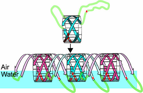
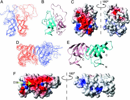
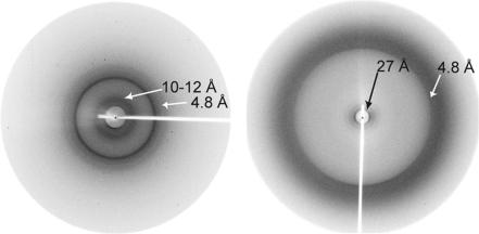
Class I hydrophobins are a unique family of fungal proteins that form a polymeric, water-repellent monolayer on the surface of structures such as spores and fruiting bodies. Similar monolayers are being discovered on an increasing range of important microorganisms. Hydrophobin monolayers are amphipathic and particularly robust, and they reverse the wettability of the surface on which they are formed. There are also significant similarities between these polymers and amyloid-like fibrils. However, structural information on these proteins and the rodlets they form has been elusive. Here, we describe the three-dimensional structure of the monomeric form of the class I hydrophobin EAS. EAS forms a beta-barrel structure punctuated by several disordered regions and displays a complete segregation of charged and hydrophobic residues on its surface. This structure is consistent with its ability to form an amphipathic polymer. By using this structure, together with data from mutagenesis and previous biophysical studies, we have been able to propose a model for the polymeric rodlet structure adopted by these proteins. X-ray fiber diffraction data from EAS rodlets are consistent with our model. Our data provide molecular insight into the nature of hydrophobin rodlet films and extend our understanding of the fibrillar beta-structures that continue to be discovered in the protein world.
Conflict of interest statement
Conflict of interest statement: No conflicts declared.
Figures

References
-
- Wösten H. A. B., van Wetter M. A., Lugones L. G., van der Mei H. C., Busscher H. J., Wessels J. G. H. Curr. Biol. 1999;9:85–88. - PubMed
-
- Wösten H. A. B., Richter M., Willey J. M. Fungal Genet. Biol. 1999;27:153–160. - PubMed
-
- Wösten H. A. B. Annu. Rev. Microbiol. 2001;55:625–646. - PubMed
-
- Wessels J. G. H. Adv. Microb. Physiol. 1997;38:1–45. - PubMed
-
- Wessels J. G. H. Annu. Rev. Phytopathol. 1994;32:413–437.
Publication types
MeSH terms
Substances
Associated data
- Actions
LinkOut - more resources
Full Text Sources
Other Literature Sources
