. 2006 Mar 14;103(11):4046-51.
doi: 10.1073/pnas.0509040103. Epub 2006 Mar 6.

Structural basis of hepatocyte growth factor/scatter factor and MET signalling

Affiliations

Structural basis of hepatocyte growth factor/scatter factor and MET signalling

Ermanno Gherardi et al. Proc Natl Acad Sci U S A. .

Abstract

The polypeptide growth factor, hepatocyte growth factor/scatter factor (HGF/SF), shares the multidomain structure and proteolytic mechanism of activation of plasminogen and other complex serine proteinases. HGF/SF, however, has no enzymatic activity. Instead, it controls the growth, morphogenesis, or migration of epithelial, endothelial, and muscle progenitor cells through the receptor tyrosine kinase MET. Using small-angle x-ray scattering and cryo-electron microscopy, we show that conversion of pro(single-chain)HGF/SF into the active two-chain form is associated with a major structural transition from a compact, closed conformation to an elongated, open one. We also report the structure of a complex between two-chain HGF/SF and the MET ectodomain (MET928) with 1:1 stoichiometry in which the N-terminal and first kringle domain of HGF/SF contact the face of the seven-blade beta-propeller domain of MET harboring the loops connecting the beta-strands b-c and d-a, whereas the C-terminal serine proteinase homology domain binds the opposite "b" face. Finally, we describe a complex with 2:2 stoichiometry between two-chain HGF/SF and a truncated form of the MET ectodomain (MET567), which is assembled around the dimerization interface seen in the crystal structure of the NK1 fragment of HGF/SF and displays the features of a functional, signaling unit. The study shows how the proteolytic mechanism of activation of the complex proteinases has been adapted to cell signaling in vertebrate organisms, offers a description of monomeric and dimeric ligand-receptor complexes, and provides a foundation to the structural basis of HGF/SF-MET signaling.


Conflict of interest statement

Conflict of interest statement: No conflicts declared.

Figures

Fig. 1.
Fig. 1.
Domain structure and biological activity of the three main proteins used in this study. (A) Domain structure. (B) SDS/PAGE under reducing conditions. (CE) Typical appearance of colonies of MDCK cells under standard culture conditions (C) or after addition of single-chain (D) or two-chain (E) HGF/SF at the concentrations indicated. sc-SF, single HGF/SF; tc-SF, two-chain HGF/SF.
Fig. 2.
Fig. 2.
Structure of single-chain and two-chain HGF/SF. (AD) Appearance of typical particles of single-chain and two-chain HGF/SF after negative staining EM (A and B) or after 3D reconstruction from CET (C and D). (E and F) The docking (45) of the crystal structure of the k2 and sp domains (blue and red, respectively) of bovine thrombin into the electron density maps of the particles shown in C and D, respectively. (G) The crystal structure of the k2-sp fragment of bovine thrombin used for docking (27) (Protein Data Bank accession code 1A0H). The k2 domain is shown in light blue, the N- and C-terminal lobes of the sp domain in dark and pale gray, respectively, and the disulphide bond connecting the α chain and the sp domain is shown in yellow. This disulphide is conserved in HGF/SF and all other members of the kringle-serine proteinase superfamily. SAXS of single-chain and two-chain HGF/SF (HJ). The scattering intensities as functions of momentum transfer [s = 4π sin(θ)/λ, where 2θ is the scattering angle and λ = 0.15 nm is the x-ray wavelength] are shown in H together with two views, at 90° from each other, of ab initio models constructed with dammin (28). Here, and in Figs. 3–5, black dots are experimental data, solid red lines are fits from ab initio models, and dashed blue lines are fits from rigid body models. (I and J) A distance-distribution plot and the rigid body modeling (29) of single-chain and two-chain HGF/SF, respectively. Individual domains are labeled as in Fig. 1, and the area of the k1 (30) and sp (21, 22) domains involved in MET binding are shown in blue and red, respectively. Images in GJ were generated with spock. sc-SF, single-chain HGF/SF; tc-SF, two-chain HGF/SF.
Fig. 3.
Fig. 3.
Structure of the MET ectodomain. (AD) Appearance of MET928 after negative staining EM (A and B) or 3D reconstruction from CET (C and D). (E) The docking of the three N-terminal domains of MET (sema, cr, and ig1) into the electron density map of the MET particle shown in C. (F and G) SAXS of the MET ectodomain. The scattering curve and an ab initio model (28) are shown in F; rigid body modeling (29) is shown in G (stalk in gray and β-propeller domain in red, except for the insertion in blade 5, which is shown in blue). sc-SF, single HGF/SF; tc-SF, two-chain HGF/SF.
Fig. 4.
Fig. 4.
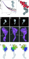
The 1:1 complex formed by two-chain HGF/SF and MET928. (A and B) SAXS of the two-chain HGF/SF-MET928 complex. (A) Scattering curve and ab initio model (28). (B) Rigid body model (29) (MET928 in gray, two-chain HGF/SF in red). (CK) CET of the two-chain HGF/SF-MET928 complex. (CE) Three views of a typical 3D reconstruction. (FH) Corresponding images after low pass filtering. (IK) Docking of the SAXS model of the two-chain HGF/SF-MET928 complex into the EM density envelope (MET β-propeller in blue; sp domain of HGF/SF in green; other HGF/SF domains in yellow); sc-SF, single-chain HGF/SF; tc-SF, two-chain HGF/SF.
Fig. 5.
Fig. 5.
The complexes formed by single-chain and two-chain HGF/SF with MET567. (A and B) SAXS of the complexes formed by single-chain and two-chain HGF/SF with MET567. (A) Scattering curves. (B) Ab initio models. In the ab initio model of the complex with single-chain HGF/SF (generated with monsa (28), MET567 is shown in yellow, and single-chain HGF/SF is shown in gray. The ab initio model of the complex of two-chain HGF/SF with MET567 was generated with dammin (28). (CE) Rigid body models of the 2:2 complex between two-chain HGF/SF and MET567 in which the dimerization interface is provided by the sp domain (C) or the n and k1 domains (D and E). In C and D, MET is shown in blue, the sp domain in red, and other HGF/SF domains in gray. In E, MET is shown in blue and the two different molecules of two-chain HGF/SF are shown in red and gray, respectively. CE were generated with spock. (F) Expression and biological activity of wild-type and mutant HGF/SF proteins. Wild-type HGF/SF and four mutants (mutA, E159A:S161A:E195A:R197A:Y198A; mutB, F82A:T83A:K85A; mutC, D123A:N127A; and mutD, V140A:I142A) were expressed transiently in Neuro2A cells. Protein levels were monitored by slot blot (Inset) and quantitated by a sandwich antibody assay. Biological activity was measured on MDCK cells (1, 4), and results were expressed as specific activity. Values are mean and standard deviation from triplicate transfections, and each mutant has been tested in at least three separate experiments. sc-SF, single-chain HGF/SF; tc-SF, two-chain HGF/SF.

References

    1. Gherardi E., Gray J., Stoker M., Perryman M., Furlong R. Proc. Natl. Acad. Sci. USA. 1989;86:5844–5848. - PMC - PubMed
    1. Nakamura T., Nishizawa T., Hagiya M., Seki T., Shimonishi M., Sugimura A., Tashiro K., Shimizu S. Nature. 1989;342:440–443. - PubMed
    1. Miyazawa K., Tsubouchi H., Naka D., Takahashi K., Okigaki M., Arakaki N., Nakayama H., Hirono S., Sakiyama O., Takahashi K., et al. Biochem. Biophys. Res. Commun. 1989;163:967–973. - PubMed
    1. Stoker M., Gherardi E., Perryman M., Gray J. Nature. 1987;327:239–242. - PubMed
    1. Weidner K. M., Arakaki N., Hartmann G., Vandekerckhove J., Weingart S., Rieder H., Fonatsch C., Tsubouchi H., Hishida T., Daikuhara Y., et al. Proc. Natl. Acad. Sci. USA. 1991;88:7001–7005. - PMC - PubMed

Publication types

MeSH terms

Substances