. 2006 Mar 14;103(11):4140-5.
doi: 10.1073/pnas.0508881103. Epub 2006 Mar 2.

Abnormal development of the olfactory bulb and reproductive system in mice lacking prokineticin receptor PKR2

Affiliations

Abnormal development of the olfactory bulb and reproductive system in mice lacking prokineticin receptor PKR2

Shun-Ichiro Matsumoto et al. Proc Natl Acad Sci U S A. .

Abstract

Prokineticins, multifunctional secreted proteins, activate two endogenous G protein-coupled receptors PKR1 and PKR2. From in situ analysis of the mouse brain, we discovered that PKR2 is predominantly expressed in the olfactory bulb (OB). To examine the role of PKR2 in the OB, we created PKR1- and PKR2-gene-disrupted mice (Pkr1(-/-) and Pkr2(-/-), respectively). Phenotypic analysis indicated that not Pkr1(-/-)but Pkr2(-/-)mice exhibited hypoplasia of the OB. This abnormality was observed in the early developmental stages of fetal OB in the Pkr2(-/-) mice. In addition, the Pkr2(-/-) mice showed severe atrophy of the reproductive system, including the testis, ovary, uterus, vagina, and mammary gland. In the Pkr2(-/-) mice, the plasma levels of testosterone and follicle-stimulating hormone were decreased, and the mRNA transcription levels of gonadotropin-releasing hormone in the hypothalamus and luteinizing hormone and follicle-stimulating hormone in the pituitary were also significantly reduced. Immunohistochemical analysis revealed that gonadotropin-releasing hormone neurons were absent in the hypothalamus in the Pkr2(-/-) mice. The phenotype of the Pkr2(-/-) mice showed similarity to the clinical features of Kallmann syndrome, a human disease characterized by association of hypogonadotropic hypogonadism and anosmia. Our current findings demonstrated that physiological activation of PKR2 is essential for normal development of the OB and sexual maturation.


Conflict of interest statement

Conflict of interest statement: No conflicts declared.

Figures
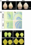
Fig. 1.
Fig. 1.
Macro- and microscopic analyses of OB from Pkr2−/− mice. (A) Macroscopic view of the male brain at 8 weeks of age from the control wild-type littermates and Pkr1−/− mice (Left), and control wild-type littermates and Pkr2−/− mice (Right). The OB size is small in the Pkr2−/− mice. (Scale bars, 5 mm.) (B) Nissl-stained sagittal sections of the male OB at 8 weeks of age from the wild-type littermates (Upper) and Pkr2−/− mice (Lower). Higher magnifications of the OB (boxes, Left) from each genotypic mouse are shown (Right), respectively. The arrow (Upper Right) indicates the glomerular layer (GL) of the OB from wild-type littermates. Note that no glomerular layer is discernible in the OB from the Pkr2−/− mice (Lower Right). [Scale bars, 0.5 mm (Left) and 0.1 mm (Right).] (C) OB morphogenesis during the embryonic stage at E14.5 (Upper Left), E16.5 (Upper Right), E18.5 (Lower Left), and P0 (Lower Right). (Scale bars, 1 mm.)
Fig. 2.
Fig. 2.
Hypoplasia of the reproductive organs in Pkr2−/− mice. (A) Macroscopic view of the testes at 8 weeks of age from the control wild-type littermates and Pkr1−/− mice (Left), control wild-type littermates and Pkr2−/− mice (Center), and the ovary and uterus (Right) at 12 weeks of age from the wild-type, Pkr1−/−, and Pkr2−/− mice. The male and female reproductive organs are small in the Pkr2−/− mice. (Scale bars, 5 mm.) (B) HE-stained sections of wild-type and Pkr2−/− testis at 20 weeks of age. Higher magnification (rectangle) shows normal Leydig cells in wild-type testis (Lower Left), whereas the mutant testis shows small Leydig cells and no spermatid (Lower Right). SPG, spermatogenic cell; SPC, spermatocytes; SPT, spermatid; LC, Leydig cells. [Scale bars, 100 μm (Upper) and 10 μm (Lower).] (C) HE-stained sections of wild-type and Pkr2−/− ovary at 20 weeks of age. A magnified region (rectangle) shows normal antral follicle (arrow) in wild-type ovary (Lower Left). A magnified region (rectangle) shows undeveloped follicles (arrows) in Pkr2−/− ovary (Lower Right). [Scale bars, 200 μm (Upper) and 50 μm (Lower).]
Fig. 3.
Fig. 3.
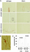
Analysis of GnRH neurons in Pkr2−/− mice. (A) Immunohistochemical study of GnRH neurons in the wild-type (Left) and Pkr2−/− mice (Right). Coronal sections of the preoptic region (Top and Middle) and median eminence (Bottom). (Scale bars, 0.1 mm.) (B) Higher magnification of the preoptic region [box (A Middle Left)] from a wild-type mouse. The arrow indicates GnRH-positive neuronal cell bodies. (Scale bar, 0.2 mm.) (C) Mean ± SD of transcriptional mRNA level of GnRH in the hypothalamus of Pkr2−/− mice. Quantitative PCR was performed to compare the mRNA level between the wild-type littermates (male, n = 6; female, n = 6) and Pkr2−/− mice (male, n = 5; female, n = 6) at 20 weeks of age. Data shown are representative of two experiments.
Fig. 4.
Fig. 4.
Abnormal morphology in the upper nasal region in Pkr2−/− mice at E12.5 and E13.5. HE-stained parasagittal sections of the upper nasal region in wild-type littermates at E12.5 (A, E, and I), Pkr2−/− mice at E12.5 (B, F, and J), wild-type littermates at E13.5 (C, G, and K) and Pkr2−/− mice at E13.5 (D, H, and L). Note that a sphere-shaped structure was observed at the region between the olfactory pit and forebrain in Pkr2−/− mice at both E12.5 and E13.5 (arrowhead in B and D). This structure is particularly evident when viewed at higher magnification (arrowhead in F, H, J, and L). OP, olfactory pit, F, forebrain. [Scale bars, 200 μm (AD), 100 μm (EH), and 50 μm (IL).]

Similar articles

Cited by

References

    1. Li M., Bullock C. M., Knauer D. J., Ehlert F. J., Zhou Q. Y. Mol. Pharmacol. 2001;59:692–698. - PubMed
    1. Bullock C. M., Li J. D., Zhou Q. Y. Mol. Pharmacol. 2004;65:582–588. - PubMed
    1. Lin D. C., Bullock C. M., Ehlert F. J., Chen J. L., Tian H., Zhou Q. Y. J. Biol. Chem. 2002;277:19276–19280. - PubMed
    1. Masuda Y., Takatsu Y., Terao Y., Kumano S., Ishibashi Y., Suenaga M., Abe M., Fukusumi S., Watanabe T., Shintani Y., et al. Biochem. Biophys. Res. Commun. 2002;293:396–402. - PubMed
    1. Soga T., Matsumoto S., Oda T., Saito T., Hiyama H., Takasaki J., Kamohara M., Ohishi T., Matsushime H., Furuichi K. Biochim. Biophys. Acta. 2002;1579:173–179. - PubMed

MeSH terms