. 2006 Apr;80(7):3180-8.
doi: 10.1128/JVI.80.7.3180-3188.2006.

The avian coronavirus infectious bronchitis virus undergoes direct low-pH-dependent fusion activation during entry into host cells

Affiliations

The avian coronavirus infectious bronchitis virus undergoes direct low-pH-dependent fusion activation during entry into host cells

Victor C Chu et al. J Virol. 2006 Apr.

Abstract

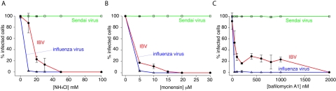
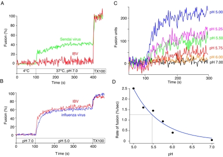
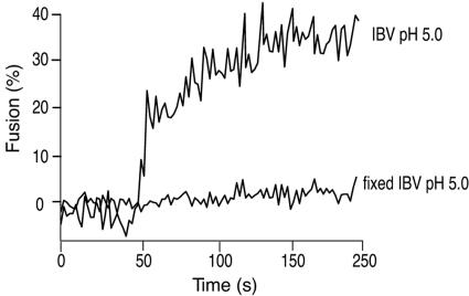
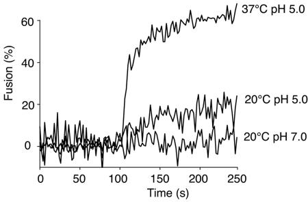
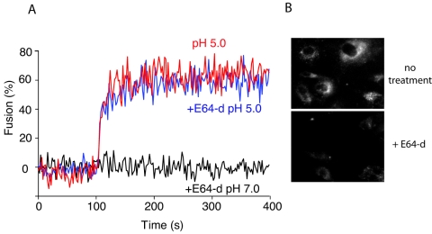
Coronaviruses are the causative agents of respiratory disease in humans and animals, including severe acute respiratory syndrome. Fusion of coronaviruses is generally thought to occur at neutral pH, although there is also evidence for a role of acidic endosomes during entry of a variety of coronaviruses. Therefore, the molecular basis of coronavirus fusion during entry into host cells remains incompletely defined. Here, we examined coronavirus-cell fusion and entry employing the avian coronavirus infectious bronchitis virus (IBV). Virus entry into cells was inhibited by acidotropic bases and by other inhibitors of pH-dependent endocytosis. We carried out fluorescence-dequenching fusion assays of R18-labeled virions and show that for IBV, coronavirus-cell fusion occurs in a low-pH-dependent manner, with a half-maximal rate of fusion occurring at pH 5.5. Fusion was reduced, but still occurred, at lower temperatures (20 degrees C). We observed no effect of inhibitors of endosomal proteases on the fusion event. These data are the first direct measure of virus-cell fusion for any coronavirus and demonstrate that the coronavirus IBV employs a direct, low-pH-dependent virus-cell fusion activation reaction. We further show that IBV was not inactivated, and fusion was unaffected, by prior exposure to pH 5.0 buffer. Virions also showed evidence of reversible conformational changes in their surface proteins, indicating that aspects of the fusion reaction may be reversible in nature.


Figures

FIG. 1.
FIG. 1.
Infection of IBV is prevented by treatment of cells with inhibitors of endosome acidification. BHK cells were treated with various concentrations of NH4Cl (A), monensin (B), or bafilomycin A1 (C) for 30 min and infected with IBV strain Beaudette, influenza virus strain A/WSN/33, or Sendai virus strain Cantell at a multiplicity of infection of 1 to 5 infectious units/cell. Infectivity was determined by immunofluorescence microscopy 8 h postinfection using anti-IBV S1 monoclonal antibody (15:88), anti-influenza NP monoclonal antibody H16 L10 4R5, or chicken anti-Sendai virus antibody. For quantification, >300 cells were scored in three independent experiments. The error bars represent the standard errors of the mean.
FIG. 2.
FIG. 2.
R18-labeled IBV virions retain infectivity in BHK cells and show no loss of S1. To determine relative infectivity, purified IBV strain Beaudette was incubated with or without R18. The protein concentration of each virus preparation was assayed via Bio-Rad protein assay, and virus infectivity was assessed by infecting BHK cells using immunofluorescence microscopy (A). To determine virus integrity, 500 ng of either R18-labeled or unlabeled IBV strain Beaudette was use to coat an ELISA plate, and the S1/S2 ratio of each sample was determined via anti-S1 (15:88) and anti-S2 (9:4) monoclonal antibody staining, followed by anti-mouse horseradish peroxidase labeling and ABTS development (B). The error bars represent the standard errors of the mean.
FIG. 3.
FIG. 3.
Fusion of IBV with host cells is low pH dependent. R18-labeled IBV strain Beaudette or Sendai virus strain Cantell was bound to BHK cells at 4°C for 60 min and then injected into a spectrofluorimeter cuvette containing 1 ml of pH 7.0 buffer at 37°C (t = 100 s) (A). Samples were monitored for fluorescence dequenching at 37°C for 300 seconds before addition of 1% Triton X-100 (final concentration) to obtain complete (100%) dequenching. (B) Similarly, R18-labeled IBV strain Beaudette or influenza virus strain A/WSN/33 was bound to BHK cells at 4°C, but samples were added to pH 7.0 buffer at 37°C (t = 0 s). At t = 100 s, the buffer pH was reduced to 5.0 and samples were monitored for fluorescence dequenching at 37°C. At t = 400 s, the final concentration of 1% Triton X-100 was added to obtain 100% dequenching. (C) Samples were treated as described for panel B under various pH conditions, and dequenching activities are shown in terms of actual fusion units. The initial rate of fusion obtained from panel C was analyzed by four-parameter exponential decay and is plotted against various pHs (D). The pH which gave the half-maximal initial rate of IBV fusion (pH1/2) is indicated.
FIG. 4.
FIG. 4.
R18-labeled IBV does not undergo significant nonspecific dye transfer. R18-labeled IBV strain Beaudette was untreated or pretreated with 0.5% paraformaldehyde (fixed IBV) before binding to BHK cells. An FdQ assay was performed on each sample as described for Fig. 4B. The pH was reduced to 5.0 to induce fusion at t = 50 s.
FIG. 5.
FIG. 5.
Limited but significant IBV fusion occurs at lower temperature. R18-labeled IBV strain Beaudette was bound to BHK cells at 4°C for 60 min and then added to a spectrofluorimeter cuvette at either 20°C or 37°C (t = 0 s). The pH was reduced to 5.0 at t = 100 s or maintained at 7.0 while FdQ activity was monitored.
FIG. 6.
FIG. 6.
Cysteine proteases are not essential for IBV fusion activation during viral entry. BHK cells were pretreated with 400 μg/ml of E64-d, and R18-labeled IBV strain Beaudette bound at 4°C for 60 min. An FdQ assay was performed as described for Fig. 4B in the presence or absence of E64-d throughout the entire experiment. Virus-cell fusion was triggered by reducing the buffer pH to 5.0 at t = 100 s. In panel B, E64-d activity was assessed using a Cathepsin L Activity Detection Kit (Calbiochem) in the presence or absence of drug treatment, according to the manufacturer's instructions.
FIG. 7.
FIG. 7.
Content mixing occurs following low-pH-induced IBV fusion at the cell surface. IBV strain Beaudette (multiplicity of infection, 5 infectious units/cell) was bound to the surfaces of BHK cells at 4°C for 60 min and was treated with 15 μM monensin to block virus entry from endosomes or was left untreated as a control. In the pH 5 pulse sample, the buffer pH was reduced to 5.0 in the presence of 15 μM of monensin for 2 min at 37°C and then replaced with 2% Dulbecco's modified Eagle's medium with monensin at 37°C for 8 h. Genome delivery and viral replication were monitored by expression of S glycoprotein via immunofluorescence microscopy. For quantification, >100 cells were scored in three independent experiments. The error bars represent the standard deviations of the mean.
FIG. 8.
FIG. 8.
Infection and fusion by IBV are not prevented by pretreatment of virions with low-pH buffer. IBV strain Beaudette, influenza virus strain A/WSN/33, and VSV strain Orsay were purified and incubated in pH 5.0 buffer for 10 min before neutralization to pH 7.0 (pH 5-pH 7) or were maintained at pH 7.0 (pH 7 only) (A). BHK cells were then infected with virus at a multiplicity of infection of 5 infectious units/cell, and infection was monitored by immunofluorescence microscopy with monoclonal antibodies anti-IBV S1 (15:88), anti-influenza virus NP H16 L10 4R5, and anti-VSV G (P5D4) after 8 h of incubation. For quantification, >100 cells were scored in three independent experiments. The error bars represent the standard deviations of the mean. (B, C, and D) IBV strain Beaudette, influenza virus strain A/WSN/33, and VSV strain Orsay were treated with either pH 5.0 or 7.0 buffer for 10 min before neutralization and then bound to BHK cells at 4°C for 60 min. FdQ assays were then performed and monitored as described for Fig. 3B, and buffer pH was reduced from pH 7.0 to 5.0 at t = 100 s.
FIG. 9.
FIG. 9.
bis-ANS labeling demonstrates S glycoprotein conformation reversibility. Purified IBV strain Beaudette, influenza virus strain A/WSN/33, and VSV strain Orsay were pretreated with pH 7.0, pH 5.0, or pH 5.0 buffer followed by neutralization to pH 7.0 at 37°C for 10 min. Then, each sample was subjected to bis-ANS binding at 37°C for 5 min before analysis by fluorimetry. The samples represent the means of five replicate wells, and the error bars represent the standard deviations of the mean.

References

    1. Beaudette, F. R., and C. R. Hudson. 1937. Cultivation of the virus of infectious bronchitis. J. Am. Vet. Med. Assoc. 90:51-60.
    1. Bosch, B. J., B. E. Martina, R. Van Der Zee, J. Lepault, B. J. Haijema, C. Versluis, A. J. Heck, R. De Groot, A. D. Osterhaus, and P. J. Rottier. 2004. Severe acute respiratory syndrome coronavirus (SARS-CoV) infection inhibition using spike protein heptad repeat-derived peptides. Proc. Natl. Acad. Sci. USA 101:8455-8460. - PMC - PubMed
    1. Bosch, B. J., R. van der Zee, C. A. de Haan, and P. J. Rottier. 2003. The coronavirus spike protein is a class I virus fusion protein: structural and functional characterization of the fusion core complex. J. Virol. 77:8801-8811. - PMC - PubMed
    1. Boulay, F., R. W. Doms, I. Wilson, and A. Helenius. 1987. The influenza hemagglutinin precursor as an acid-sensitive probe of the biosynthetic pathway. EMBO J. 6:2643-2650. - PMC - PubMed
    1. Cavanagh, D. 2005. Coronaviridae: a review of coronaviruses and toroviruses, p. 1-54. In A. Schmidt, M. H. Wolff, and O. Weber (ed.), Coronaviruses with special emphasis on first insights concerning SARS. Birkhauser Verlag, Basel, Switzerland.

Publication types

Substances

LinkOut - more resources