. 2006 Apr;80(7):3249-58.
doi: 10.1128/JVI.80.7.3249-3258.2006.

Membrane-anchored inhibitory peptides capture human immunodeficiency virus type 1 gp41 conformations that engage the target membrane prior to fusion

Affiliations

Membrane-anchored inhibitory peptides capture human immunodeficiency virus type 1 gp41 conformations that engage the target membrane prior to fusion

Gregory B Melikyan et al. J Virol. 2006 Apr.

Abstract

Soluble peptides derived from the C-terminal heptad repeat domain of human immunodeficiency virus type 1 (HIV-1) gp41 are potent inhibitors of HIV-1 entry and gp41-induced fusion. Target membrane-anchored variants of these peptides have been shown to retain inhibitory activity. Both soluble and membrane-anchored C peptides (MACs) are thought to block fusion by binding to the N-terminal coiled coil domain of gp41 and preventing formation of the final six-helix bundle structure. However, interactions of target MACs with gp41 must be restricted to a subset of trimers that have their hydrophobic fusion peptides inserted into the target membrane. This unique feature of MACs was used to identify the intermediate step of fusion at which gp41 engaged the target membrane. Fusion between HIV envelope-expressing effector cells and target cells was measured by fluorescence microscopy. Expression of MACs in target cells led to less than twofold reduction in the extent of fusion. However, when reaction was first arrested by adding lysolipids that disfavored membrane merger, and the lipids were subsequently removed by washing, control cells supported fusion, whereas those that expressed MACs did not. The drastically improved potency of MACs implies that, at lipid-arrested stage, gp41 bridges the viral and target cell membranes and therefore more optimally binds the membrane-anchored peptides. Experimental demonstration of this intermediate shows that, similar to fusion induced by many other viral glycoproteins, engaging the target membrane by HIV-1 gp41 permits coupling between six-helix bundle formation and membrane merger.


Figures

FIG. 1.
FIG. 1.
A model of HIV Env-induced fusion and its inhibition by MACs. Intermediate conformations of gp41 are shown without gp120 for visual clarity. The C- and N-terminal heptad repeat domains are represented by blue and red cylinders, respectively. Fusion peptides are shown by arrows. Upper panel illustrates the proposed mechanism of inhibition by the tMACs, while the lower panel depicts the proposed mode of action of eMACs. The red X indicates the putative fusion step blocked by the peptides.
FIG. 2.
FIG. 2.
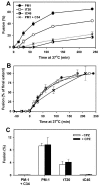
The kinetics of HIV-1 Env-induced fusion to target PM-1 cells that do not express (filled circles) or express tT20 or tC46 (open and half-filled circles, respectively). (A) 293T cells transiently transfected with X4-tropic HIV Env (effector cells) were labeled with calcein, mixed with target cells loaded with CMAC, and coincubated at 37°C for varied times. Fusion was stopped at the indicated time intervals by adding 1 μM C34 peptide, and the extent of fusion was determined, as described in Materials and Methods. In control experiments, PM-1 cells were fused to effector cells in the presence of 75 nM of soluble C34 peptide (crosses). (B) Data from panel A were normalized to the extent of fusion after a 4-h incubation, and the curves were aligned at the onset of content mixing by subtracting the appropriate lag times to fusion. (C) The effect of chlorpromazine treatment (CPZ, filled bars) on fusion to target cells expressing (tT20 and tC46) or not expressing (PM-1) tMACs. Following a 30-min coincubation at 37°C, cells were treated with 0.5 mM CPZ in phosphate-buffered saline (PBS) for 1 min at room temperature, washed twice, and examined under a microscope. Control cells (open bars) were treated with PBS. Note that target cells were loaded with CMFDA instead of calcein to minimize the dye leakage caused by CPZ. To test whether hemifusion occurred in the presence of free C34, 1 μM C34 was added to the mixture of effector and target (PM-1) cells at the time of coculture (first column). Unless stated otherwise, the experimental points are means and standard errors of results from at least four independent experiments performed in duplicate.
FIG. 3.
FIG. 3.
Synergy between the inhibitory effects of tT20 and AMD3100. (A) Fusion to PM-1 and tT20 cells (open and filled circles, respectively) in the presence of varied concentrations of AMD3100. Cells were coincubated for 2 h at 37°C. (B) Inhibition of fusion by AMD3100 added at TAS. Effector cells were preincubated with PM-1 (open circles) or tT20 (filled circles) cells for 2.5 h at 23°C to create the TAS, treated with indicated concentrations of AM3100 (5 min at 23°C), and warmed to 37°C for 1 h. AMD3100 used for experiments in panels A and B was obtained through NIAID ARRRP. (C) PM-1 and tT20 cells (open and filled circles, respectively) were coincubated at 23°C with Env-expressing cells for 1, 2.5, or 3.5 h, followed by addition of 10 μM AMD3100 (obtained from Sigma) and further incubation for 1 h at 37°C. The acquisition of resistance to AMD3100 was expressed as the ratio of fusion in the presence of the drug to that in the absence of the drug. Experimental points are means ± standard errors of the means of results from two independent experiments performed in duplicate. (D) Kinetics of fusion to PM-1 and tT20 cells in the absence (open and filled circles, respectively) and in the presence (open and filled triangles) of 190 nM AMD3100 added at the beginning of cell coincubation at 37°C.
FIG. 4.
FIG. 4.
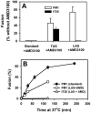
Inhibitory potency of tMACs in a standard protocol and after arresting fusion at intermediate stages upstream of membrane merger. (A) HIV-1 Env-expressing 293T cells were fused to PM-1 (open bars) or tT20 (filled bars) cells. Fusion from the TAS (second column) and LAS (third column) was triggered in the presence of 20 μM AMD3100 (from NIAID ARRRP) to prevent dye transfer between cells that did not form ternary complexes at these stages (for details, see Materials and Methods). The extent of fusion was normalized to that obtained for PM-1 cells in the absence of AMD3100. (B) Kinetics of fusion to PM-1 cells (open circles) and tT20 cells (open triangles) induced by removing LPC (after arresting fusion at LAS) in the presence of 20 μM AMD3100 (NIAID ARRRP). The kinetics of fusion to PM-1 cells in a standard protocol is shown for comparison (filled circles, replotted from Fig. 2A).
FIG. 5.
FIG. 5.
The inhibitory activity of the membrane-anchored C46 expressed in effector (eC46) or in target (tC46) cells. (A) Fusion between 293T cells (E) expressing Env (loaded with green fluorescent dye, CMFDA) and 293T cells (T) transfected with CD4 (loaded with blue dye, CMAC) was quantified following 2 h of coculture at 37°C (first bar). The effect of C46 was evaluated by fusing target cells to effector cells coexpressing Env and C46 (eC46+T, second bar) or, alternatively, by fusing cells expressing Env to target cells that coexpressed CD4 and C46 (E+tC46, third bar). The extent of dye transfer was normalized to fusion between E/T cell pairs lacking the inhibitory peptide (first bar). (B) Dye transfer between 293T cells expressing Env and eC46 and target 293T cells expressing CD4 was measured in a standard protocol, after creating the TAS or LAS (eC46+T, filled bars). Fusion from the TAS and LAS was triggered in the presence of 20 μM AMD3100 (NIAID ARRRP). The data were normalized to the extent of fusion between target and effector cells lacking the inhibitory peptide. For comparison, the normalized extents of fusion between effector and tT20-expressing PM-1 cells are shown for all three fusion protocols (open bars).
FIG. 6.
FIG. 6.
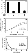
Inhibitory activity of C46 anchored to effector cell membrane (eC46). (A) Effector cells either not expressing (first and second bar) or expressing (third and fourth bar) the eC46 were pretreated with 7 μg/ml sCD4 (10 min, 37°C), washed twice, and coincubated with PM-1 cells for 2 h at 37°C. The effect of sCD4 on fusion was quantified by normalizing the fraction of fused cells to that in the absence of the soluble receptor. (B) Effector cells lacking (circles) or expressing (triangles) eC46 were coincubated with PM-1 cells at 23°C for varied times, exposed to 10 μM AMD3100, and warmed to 37°C. The acquisition of resistance to AMD3100 is expressed as the ratio of fusion in the presence of AMD3100 to that in the absence of the drug. Experimental points are means ± standard errors of the means of results from two independent experiments performed in duplicate. (C) Dose dependences of inhibition of fusion between control cells lacking MACs (E+PM1, open circles), between eC46-expressing and PM-1 cells (eC46+PM1, open triangles), or between effector and tT20-expressing cells (E+tT20, filled circles). The data were normalized to fusion in the absence of the drug. AMD3100 used in the experiments for which results are shown in Fig. 6 was purchased from Sigma.

Similar articles

Cited by

References

    1. Abrahamyan, L. G., R. M. Markosyan, J. P. Moore, F. S. Cohen, and G. B. Melikyan. 2003. Human immunodeficiency virus type 1 Env with an intersubunit disulfide bond engages coreceptors but requires bond reduction after engagement to induce fusion. J. Virol. 77:5829-5836. - PMC - PubMed
    1. Abrahamyan, L. G., S. R. Mkrtchyan, J. Binley, M. Lu, G. B. Melikyan, and F. S. Cohen. 2005. The cytoplasmic tail slows the folding of human immunodeficiency virus type 1 Env from a late prebundle configuration into the six-helix bundle. J. Virol. 79:106-115. - PMC - PubMed
    1. Andre, S., B. Seed, J. Eberle, W. Schraut, A. Bultmann, and J. Haas. 1998. Increased immune response elicited by DNA vaccination with a synthetic gp120 sequence with optimized codon usage. J. Virol. 72:1497-1503. - PMC - PubMed
    1. Binley, J. M., C. S. Cayanan, C. Wiley, N. Schulke, W. C. Olson, and D. R. Burton. 2003. Redox-triggered infection by disulfide-shackled human immunodeficiency virus type 1 pseudovirions. J. Virol. 77:5678-5684. - PMC - PubMed
    1. Binley, J. M., R. W. Sanders, B. Clas, N. Schuelke, A. Master, Y. Guo, F. Kajumo, D. J. Anselma, P. J. Maddon, W. C. Olson, and J. P. Moore. 2000. A recombinant human immunodeficiency virus type 1 envelope glycoprotein complex stabilized by an intermolecular disulfide bond between the gp120 and gp41 subunits is an antigenic mimic of the trimeric virion-associated structure. J. Virol. 74:627-643. - PMC - PubMed

Publication types

MeSH terms

LinkOut - more resources