. 2006 Apr;80(7):3341-8.
doi: 10.1128/JVI.80.7.3341-3348.2006.

Protein kinase B/Akt is present in activated form throughout the entire replicative cycle of deltaU(S)3 mutant virus but only at early times after infection with wild-type herpes simplex virus 1

Affiliations

Protein kinase B/Akt is present in activated form throughout the entire replicative cycle of deltaU(S)3 mutant virus but only at early times after infection with wild-type herpes simplex virus 1

Luca Benetti et al. J Virol. 2006 Apr.

Abstract

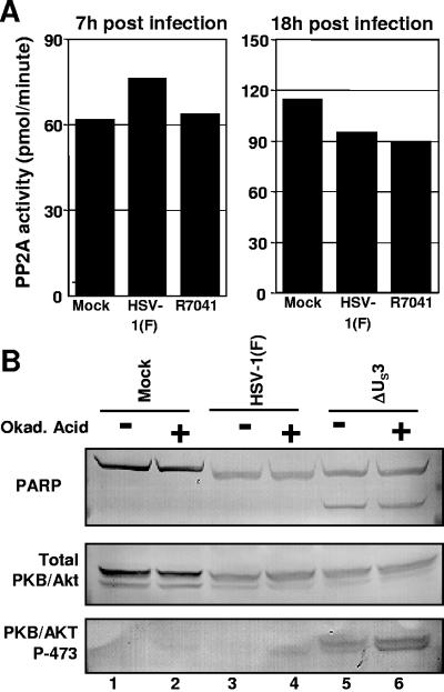
The product of the herpes simplex virus 1 (HSV-1) US3 gene is a multifunctional serine-threonine protein kinase that can block apoptosis induced by proapoptotic cellular proteins, exogenous agents, or replication-defective viruses. Earlier studies showed that the U(S)3 kinase activates and functionally overlaps cellular protein kinase A (PKA). In this study we examined the status of phosphatidylinositol 3-kinase [PI3K] and of its effector, protein kinase B/Akt (PKB/Akt), a component of a major pathway of mammalian antiapoptotic signaling systems. We report the following. (i) Infection of target cells with HSV-1 induces transient phosphorylation of serine 473 of PKB/Akt early in infection, with a mechanism that is dependent on PI3K. Inhibition of PI3K induced apoptosis in mock-infected or deltaU(S)3 mutant-virus-infected but not in wild-type-virus-infected cells and reduced the accumulation of specific viral gene products, including the U(S)3 protein kinase, but had a marginal effect on virus yields. (ii) At later times after infection, the total amounts of PKB/Akt decreased and phosphorylated PKB/Akt forms disappeared in a U(S)3-dependent and protein phosphatase 2A-independent manner. (iii) Activation of PKA by forskolin did not mediate significant dephosphorylation of PKB/Akt. Our results are consistent with the model that PKB/Akt is activated early in infection and acts to block apoptosis in infected cells prior to the accumulation of U(S)3 protein kinase and that it persists and continues to function as an antiapoptotic protein in the absence of U(S)3 but becomes redundant or even inimical once U(S)3 protein kinase accumulates in effective amounts.


Figures

FIG. 1.
FIG. 1.
Photographs of electrophoretically separated cell lysates reacted with antibody against total PKB/Akt, phosphorylated PKB/Akt serine 473 or threonine 308, US3, or β-actin. HEp-2 cells were mock infected or exposed to 10 PFU of HSV-1(F), ΔUS3, or ΔICP4 per cell and maintained in DMEM supplemented with 5% NBCS. The cells were harvested at the time points shown, solubilized, and normalized for protein content. Equal amounts of protein lysates (120 μg/lane) were subjected to electrophoresis on a denaturing gel, transferred to a nitrocellulose sheet, and reacted with antibodies as indicated.
FIG. 2.
FIG. 2.
Proteasomal inhibitors do not affect the accumulation of total PKB/Akt in infected HEp2 cells. (A) Cells were mock infected or exposed to 10 PFU of HSV-1(F) or ΔUS3 mutant virus per cell, maintained for 6.5 h in DMEM supplemented with 5% NBCS, and treated with 50 μM MG132 for 2.5 h. (B) HEp-2 cells were mock infected or exposed to 10 PFU of HSV-1(F) or ΔUS3 or ΔICP4 mutant virus per cell, maintained for 5 h in DMEM supplemented with 5% NBCS, and treated with 10 μM lactacystin for 2.5 h. The cells were then harvested and solubilized. Equal amounts of protein lysates (120 μg/lane) were subjected to electrophoresis and subsequent immunoblotting with the indicated antibodies.
FIG. 3.
FIG. 3.
Role of PI(3)K pathway in PKB/Akt phosphorylation and herpesvirus gene expression. (A) Replicate cultures of HEp-2 cells were mock infected or exposed to 10 PFU of HSV-1(F) or ΔUS3 mutant virus per cell; after 7 h, cells were exposed to EGF (100 ng/ml) for 20 min, harvested, solubilized, and normalized for protein content. Equal amounts of protein lysates (120 μg/lane) were subjected to electrophoresis on a denaturing gel, transferred to a nitrocellulose sheet, and reacted with the indicated antibodies. (B) HEp-2 cells were mock infected or incubated with HSV-1(F) for 1 h and then exposed to the PI(3)K inhibitor LY294002 (75 μM) for 2 h. The cells were then treated with EGF (100 ng/ml) for an additional 30 min, harvested, and solubilized. Equal amounts of protein lysates (120 μg/lane) were subjected to electrophoresis and immunoblotting with the indicated antibodies. (C) Replicate cultures of HEp-2 cells were mock infected or exposed to 10 PFU of HSV-1(F) or ΔUS3 or ΔICP4 mutant virus per cell and then exposed to LY294002 (75 μM) solubilized in dimethyl sulfoxide or to an equivalent amount of dimethyl sulfoxide added to the medium. The cells were harvested at 3.5 h after the infection and processed as described above. While 120 μg/lane of protein lysates was used for immunoblotting with antibodies against total and phosphorylated PKB/Akt, 60 μg/lane was used for immunoblotting with antibodies against viral proteins.
FIG. 4.
FIG. 4.
Effect of PI(3)K inhibition on infected-cell viability. Replicate cultures of HEp-2 cells were mock infected or exposed to 10 PFU of HSV-1(F) or ΔUS3 or ΔICP4 mutant virus per cell, maintained for 5 h in DMEM supplemented with 5% NBCS, and then exposed to the PI(3)K inhibitor LY294002 (75 μM) or medium containing dimethyl sulfoxide as described above. The cells were harvested at 18 h after infection, solubilized, and processed as described above for immunoblotting with antibody directed against PARP (A) or tested for DEVDase activity (B). The results for the DEVDase activity assays are indicated as increase (-fold) above activity of untreated, mock-infected cells.
FIG. 5.
FIG. 5.
(A) Assays of PP2A activity in HSV-1(F)-infected cells. Replicate cultures of HEp-2 cells were either mock infected or exposed to HSV-1(F) or the ΔUS3 mutant virus. At the indicated time points, cells were harvested and lysed with a Dounce homogenizer. The PP2A was precipitated from the lysates with the antibody to the catalytic subunit of PP2A. The immune-precipitated PP2A was then assayed for phosphatase activity, measured as release of phosphate groups in the supernatant fluid from a synthetic PP2A substrate as described in Materials and Methods. (B) Effect of okadaic acid treatment on the accumulation of phospho-Akt in HSV-1-infected cells. Replicate cultures of HEp-2 cells were either mock infected or exposed to 10 PFU of HSV-1(F) or ΔUS3 mutant virus per cell for 1 h and maintained in DMEM supplemented with 5% NBCS for 11 h. At that time the cells were mock treated or exposed to okadaic acid (50 ng/ml) for an additional 7 h. The cells were then harvested and solubilized, and the lysates were normalized for protein content: equal amounts of protein lysates were subjected to electrophoresis on a denaturing gel and reacted with antibody against either PARP, total PKB/Akt, or PKB/Akt P-473.
FIG. 6.
FIG. 6.
Effect of forskolin treatment on the accumulation of phospho-Akt. For lanes 1 to 16, HEp-2 cells were either mock infected or exposed to 10 PFU of HSV-1(F) or ΔUS3 per cell for 1 h. The cells were then exposed to medium containing forskolin (100 μg/ml) initially dissolved in ethanol or medium containing an equivalent amount of ethanol for 2.5 h. For lanes 17 to 24, the cells were maintained in DMEM supplemented with 5% NBCS for 3 h, then treated with forskolin as described above, and harvested at 17 h postinfection. The cells were solubilized, and equal amounts of protein lysates were subjected to electrophoresis on a denaturing gel, and reacted with antibody against PARP or against either total PKB/Akt or PKB/Akt P-473.

References

    1. Ackermann, M., D. K. Braun, L. Pereira, and B. Roizman. 1984. Characterization of herpes simplex virus 1 alpha proteins 0, 4, and 27 with monoclonal antibodies. J. Virol. 52:108-118. - PMC - PubMed
    1. Ahmed, N. N., H. Leighton Grimes, A. Bellacosa, T. O. Chan, and P. N. Tsichilis. 1997. Transduction of interleukin-2 antiapoptotic and proliferative signals via Akt protein kinase. Proc. Natl. Acad. Sci. USA 94:3627-3632. - PMC - PubMed
    1. Alessi, D. R., M. Andjelkovic, B. Caudwell, P. Cron, N. Morrice, P. Cohen, and B. A. Hemmings. 1996. Mechanism of activation of protein kinase B by insulin and IGF-1. EMBO J. 15:6541-6551. - PMC - PubMed
    1. Benetti, L., and B. Roizman. 2004. Herpes simplex virus protein kinase US3 activates and functionally overlaps protein kinase A to block apoptosis. Proc. Natl. Acad. Sci. USA 101:9411-9416. - PMC - PubMed
    1. Benetti, L., J. Munger, and B. Roizman. 2003. The herpes simplex virus 1 US3 protein kinase blocks caspase dependent double cleavage and activation of the proapoptotic protein BAD. J. Virol. 77:6567-6573. - PMC - PubMed

Publication types

MeSH terms

Substances

LinkOut - more resources