Comparative Study
. 2006 Apr;80(7):3416-27.
doi: 10.1128/JVI.80.7.3416-3427.2006.

A simian virus 5 (SV5) P/V mutant is less cytopathic than wild-type SV5 in human dendritic cells and is a more effective activator of dendritic cell maturation and function

Affiliations
Comparative Study

A simian virus 5 (SV5) P/V mutant is less cytopathic than wild-type SV5 in human dendritic cells and is a more effective activator of dendritic cell maturation and function

Subhashini Arimilli et al. J Virol. 2006 Apr.

Abstract

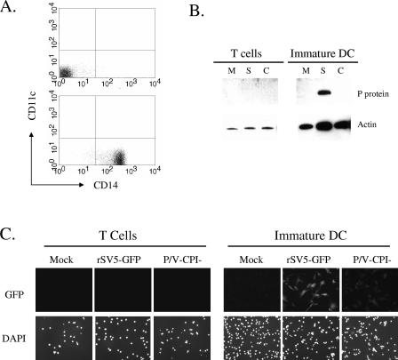
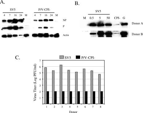
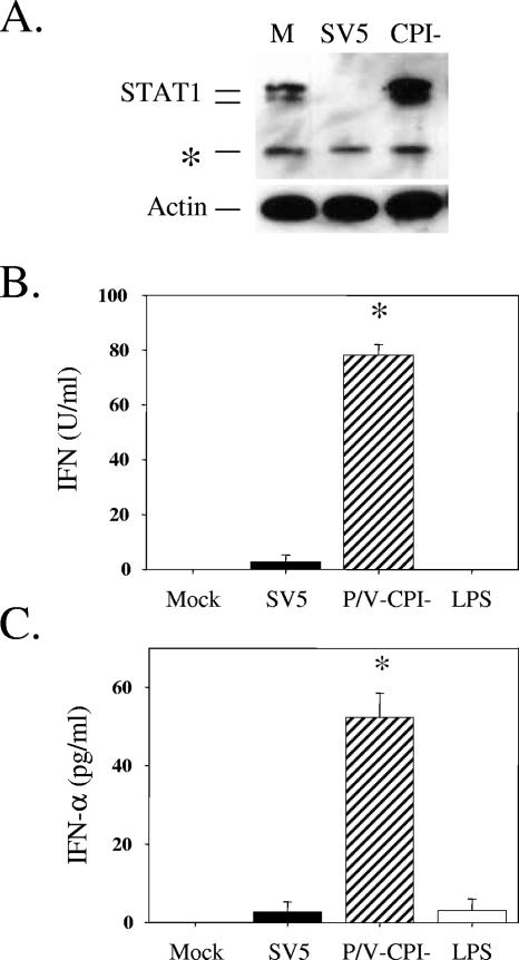
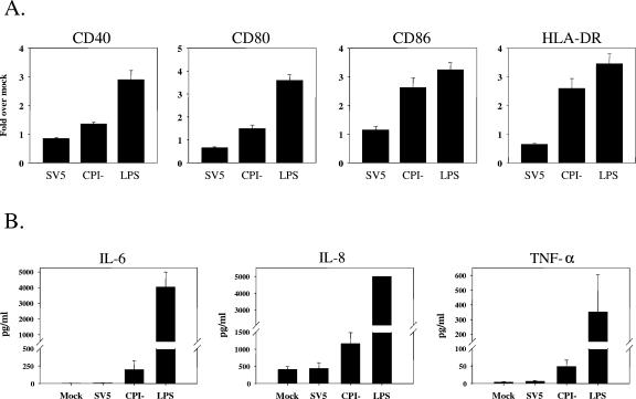
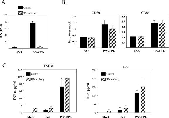
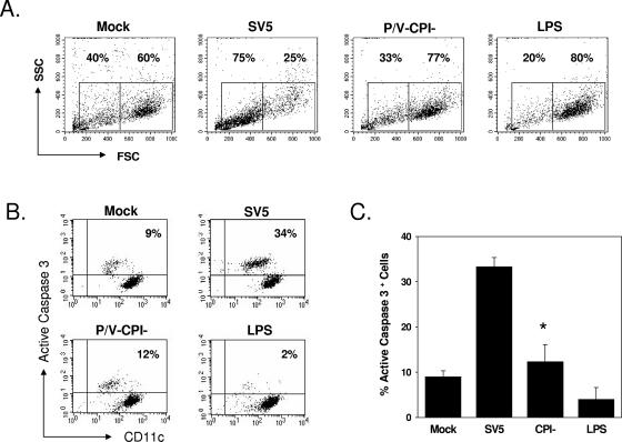
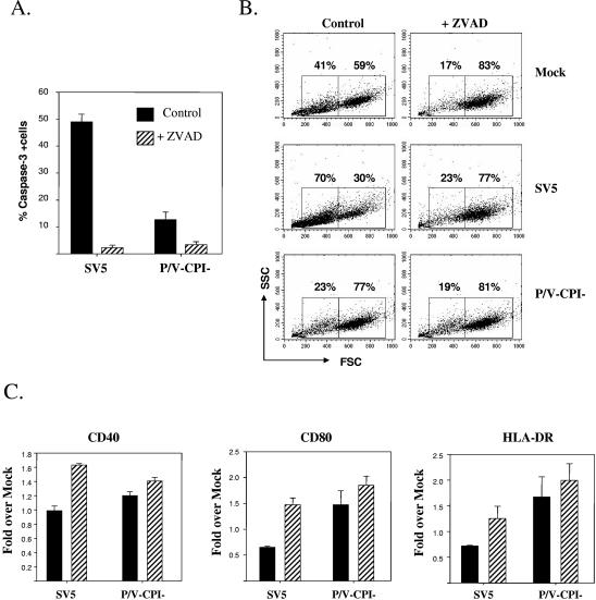
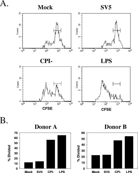
Human epithelial cells infected with the parainfluenza virus simian virus 5 (SV5) show minimal activation of host cell interferon (IFN), cytokine, and cell death pathways. In contrast, a recombinant SV5 P/V gene mutant (rSV5-P/V-CPI-) overexpresses viral gene products and is a potent inducer of IFN, proinflammatory cytokines, and apoptosis in these cells. In this study, we have compared the outcomes of wild-type (WT) SV5 and rSV5-P/V-CPI- infections of primary human dendritic cells (DC), important antigen-presenting cells for initiating adaptive immune responses. We have tested the hypothesis that a P/V mutant which activates host antiviral responses will be a more potent inducer of DC maturation and function than WT rSV5, which suppresses host cell responses. Infection of peripheral blood mononuclear cell-derived immature DC with WT rSV5 resulted in high levels of viral protein and progeny virus but very little increase in cell surface costimulatory molecules or secretion of IFN and proinflammatory cytokines. In contrast, immature DC infected with the rSV5-P/V-CPI- mutant produced only low levels of viral protein and progeny virus, but these infected cells were induced to secrete IFN-alpha and other cytokines and showed elevated levels of maturation markers. Unexpectedly, DC infected with WT rSV5 showed extensive cytopathic effects and increased levels of active caspase-3, while infection of DC with the P/V mutant was largely noncytopathic. In mixed-culture assays, WT rSV5-infected DC were impaired in the ability to stimulate proliferation of autologous CD4+ T cells, whereas DC infected with the P/V mutant were very effective at activating T-cell proliferation. The addition of a pancaspase inhibitor to DC infected with WT rSV5 reduced cytopathic effects and resulted in higher surface expression levels of maturation markers. Our finding that the SV5 P/V mutant has both a reduced cytopathic effect in human DC compared to WT SV5 and an enhanced ability to induce DC function has implications for the rational design of novel recombinant paramyxovirus vectors based on engineered mutations in the viral P/V gene.


Figures

FIG. 1.
FIG. 1.
SV5 infection of primary human T cells and immature DC. (A) PBMC were separated by selection with CD14 beads, and the unselected (top panel) or selected (bottom panel) populations were analyzed by flow cytometry for surface CD14 and CD11c expression. (B) T cells or immature DC were mock infected (lanes M) or infected at an MOI of 5 with rSV5-GFP (lanes S) or P/V-CPI (lanes C). At 24 h p.i., cell lysates were prepared and analyzed by Western blotting for SV5 P protein or cellular actin. (C) T cells or immature DC were mock infected or infected at an MOI of 5 with rSV5-GFP or P/V-CPI and examined at 24 h p.i. by microscopy for nuclei (DAPI panels) or GFP. Panels represent equivalent exposure times.
FIG. 2.
FIG. 2.
DC infected by the SV5 P/V mutant show low levels of viral protein expression and low virus yields. (A) Time course of protein expression. DC isolated from a representative donor were mock infected (lanes M) or infected at an MOI of 5 with rSV5-GFP or the P/V-CPI mutant. Protein extracts were collected at the indicated times (h) p.i. and analyzed by Western blotting for SV5 NP and P or cellular actin. (B) Dose-dependent protein expression. Immature DC isolated from two representative donors were mock infected (lane M), infected at the indicated MOI with rSV5, or infected at an MOI of 5 with rSV5-P/V-CPI (lane CPI) or WT rSV5-GFP (lane G). Protein extracts were analyzed by Western blotting with a polyclonal antiserum specific for the SV5 P protein. (C) Virus yields. DC isolated from eight individual donors were infected at an MOI of 5 with WT rSV5 or with the P/V-CPI mutant, and virus yields at 24 h p.i. were determined by a plaque assay.
FIG. 3.
FIG. 3.
STAT1 and IFN levels following infection of DC by WT rSV5 and the P/V mutant. (A) STAT1 levels. Immature DC from a representative donor were mock infected (lane M) or infected at an MOI of 5 with rSV5-GFP or the P/V-CPI mutant virus. Protein extracts were analyzed by Western blotting with a polyclonal antiserum specific for STAT1 or cellular actin. *, cross-reactive host protein. (B and C) Type I IFN levels. DC isolated from individual donors were mock infected, infected with rSV5 or the P/V-CPI mutant at an MOI of 5, or treated with LPS (200 ng/ml). At 24 h p.i., supernatants were analyzed by a VSV challenge assay for levels of type I IFN (B) or by ELISA for levels of IFN-α (C). Data are the mean values from three donors, with standard deviations indicated by bars. *, significantly different from WT SV5-infected samples.
FIG. 4.
FIG. 4.
Increases in maturation markers following infection of immature DC with the P/V-CPI mutant but not with WT rSV5. DC from individual donors were mock infected, infected at an MOI of 5 with WT rSV5 or the P/V-CPI mutant, or treated with LPS. After 24 h, cells were stained with antibodies to the indicated maturation marker proteins and analyzed by flow cytometry (A). Mean fluorescence intensities (with standard errors) are expressed relative to those of mock samples and are from four independent experiments with DC from four donors. The upregulation of costimulatory molecules following CPI mutant infection compared to that after SV5 infection was statistically significant, with the following P values: for CD40, P = 0.00034; for CD80, P = 0.0069; for CD86, P = 0.0086; for HLA-DR, P = 0.01. Alternatively, cell-free supernatants were assayed by a cytometric bead array for the indicated cytokines (B). Values are means ± standard errors of three independent experiments with DC from four donors.
FIG. 5.
FIG. 5.
Role of type I IFN secretion in DC maturation following infection with WT and P/V mutant SV5. DC from individual donors were mock infected or infected at an MOI of 5 with WT rSV5 or the P/V-CPI mutant and incubated with (hatched bars) or without (black bars) a mixture of neutralizing antibodies against IFN-α and IFN-β. After 24 h, samples were harvested, and supernatants were analyzed by a VSV challenge assay for levels of type I IFN (A). Alternatively, cells were stained with antibodies to the indicated maturation marker proteins and analyzed by flow cytometry (B). Mean fluorescence intensities (with standard errors) are expressed relative to those of mock samples and are from four independent experiments with cells from four donors. Cell-free supernatants were also assayed by a cytometric bead array for TNF-α and IL-6 (C). Values are means ± standard errors of three independent experiments with DC from three donors.
FIG. 6.
FIG. 6.
Increased cytopathic effect and active caspase-3 following infection of DC with WT rSV5 but not with the P/V-CPI mutant. DC from individual donors were mock infected, infected at an MOI of 5 with WT rSV5 or the P/V-CPI mutant, or treated with LPS (200 ng/ml). At 24 h p.i., cells were analyzed by flow cytometry for intracellular levels of active caspase-3. Panel A shows results from a representative experiment plotting forward and side scatter. Panel B shows results from a representative experiment plotting levels of staining with FITC-anti-caspase-3 antibody (y axis) versus staining with phycoerythrin-Cy5-anti-CD11c antibody (x axis). Panel C shows the mean percentages of cells with elevated caspase-3 staining from four individual donors. *, P value of 0.026 between SV5- and CPI mutant-infected samples.
FIG. 7.
FIG. 7.
Z-VAD-FMK treatment reduces active caspase-3 and cytopathic effects in SV5-infected DC. (A) Active caspase-3 levels. DC were infected at an MOI of 5 with WT rSV5 or the P/V-CPI mutant and incubated for 24 h with or without 100 μM Z-VAD-FMK (pancaspase inhibitor) before analysis of caspase-3 levels by flow cytometry. Data are the means ± standard errors from three individual experiments with three donors. The values for SV5-infected DC with and without Z-VAD-FMK treatment were significantly different (P = 0.0001) by a paired t test. (B and C) Cytopathic effect and maturation markers. Mock-infected or infected DC were treated as described for panel A and analyzed by flow cytometry for changes in forward and side scatter (B). In addition, cells were analyzed for cell surface maturation marker levels by flow cytometry (C). Mean fluorescence intensities (with standard errors) are expressed relative to those of mock samples and are from four independent experiments with four donors.
FIG. 8.
FIG. 8.
DC infected with the P/V mutant, but not with WT rSV5, are effective activators of T-cell proliferation. DC were mock treated, infected at an MOI of 5 with WT SV5 or the P/V-CPI mutant, or treated with LPS (200 ng/ml) for 24 h before being cocultured with CFSE-labeled autologous T cells in the presence of 0.01 ng/ml TSST-1 superantigen. After 3 days, cells were stained with TCR Vβ2-APC antibodies (y axis), and the proliferation of CD4+ cells was measured by changes in CFSE intensity (x axis). (A) Representative CFSE staining results for an individual donor's DC. (B) Percentages of T cells undergoing division (% divided) for samples from two individual donors.

Similar articles

Cited by

References

    1. Andrejeva, J., D. F. Young, S. Goodbourn, and R. E. Randall. 2002. Degradation of STAT1 and STAT2 by the V proteins of SV5 and human parainfluenza virus type 2, respectively: consequences for virus replication in the presence of alpha/beta and gamma interferons. J. Virol. 76:2159-2167. - PMC - PubMed
    1. Andrejeva, J., K. S. Childs, D. F. Young, T. S. Carlos, N. Stock, S. Goodbourn, and R. E. Randall. 2004. The V proteins of paramyxoviruses bind the IFN-inducible RNA helicase Mda-5 and inhibit its activation of the IFN-beta promoter. Proc. Natl. Acad. Sci. USA 101:17264-17269. - PMC - PubMed
    1. Asselin-Paturel, C., and G. Trinchieri. 2005. Production of type I interferons: plasmacytoid dendritic cells and beyond. J. Exp. Med. 202:461-465. - PMC - PubMed
    1. Baize, S., J. Kaplon, C. Faure, D. Pannetier, M. C. Georges-Courbot, and V. Deubel. 2004. Lassa virus infection of human dendritic cells and macrophages is productive but fails to activate cells. J. Immunol. 172:2861-2869. - PubMed
    1. Banchereau, J., F. Briere, C. Caux, J. Davoust, S. Lebecque, Y. J. Liu, B. Pulendran, and K. Palucka. 2000. Immunobiology of dendritic cells. Annu. Rev. Immunol. 18:767-811. - PubMed

Publication types

MeSH terms