. 2006 Apr;80(7):3445-58.
doi: 10.1128/JVI.80.7.3445-3458.2006.

Kaposi's sarcoma-associated herpesvirus-encoded latency-associated nuclear antigen modulates K1 expression through its cis-acting elements within the terminal repeats

Affiliations

Kaposi's sarcoma-associated herpesvirus-encoded latency-associated nuclear antigen modulates K1 expression through its cis-acting elements within the terminal repeats

Subhash C Verma et al. J Virol. 2006 Apr.

Abstract

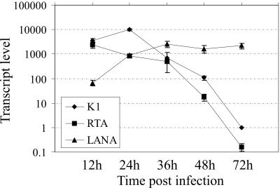
K1 is the first open reading frame encoded by Kaposi's sarcoma-associated herpesvirus (KSHV) and lies positionally to the immediate right of the terminal repeats. K1 is a transmembrane glycoprotein having a functional immunoreceptor tyrosine-based activation motif (ITAM) capable of activating B-cell receptor signaling. K1 is expressed mostly during the lytic cycle of the virus and its promoter lies within the terminal repeat which contains the binding sites for latency-associated nuclear antigen (LANA). The K1 promoter (K1p) having LANA binding sites assayed by reporter assay demonstrated that LANA is capable of down-regulating K1 promoter transcriptional activity. However, the KSHV replication transcription activator RTA up-regulates K1p transcriptional activity. The promoter deleted of LANA binding sites showed loss in LANA-mediated down-regulation but was unaffected for RTA-mediated up-regulation. Increasing amounts of RTA rescued LANA-mediated repression of K1p transcriptional activity in cotransfection experiments. Reporter assay data suggest that LANA binding to its cognate sequence is critical for LANA-mediated repression of K1p as a LANA construct lacking the DNA binding domain was unable to repress K1p transcription. Additionally, KSHV primary infection experiments suggest that K1 is expressed during early infection but is repressed on the establishment of latency and so follows an expression profile similar to that of RTA during infection. Analysis of the promoter sequence revealed the presence of Oct-1 transcription factor binding sites within the -116 to +76 region. Mutational analysis of the Oct-1 sites abolished RTA-mediated transcriptional activation, suggesting that RTA up-regulates K1p transcription through binding to this transcription factor.


Figures

FIG. 1.
FIG. 1.
LANA down-regulates K1 promoter transcriptional activity. A. Strategy for cloning K1p into pGL3B. The NotI-BamHI fragment of cosmid Z6 containing K1 and the terminal repeat was excised and cloned into BSpuro at the indicated sites, generating pBSpuroB. The coding sequence of K1 was deleted by digesting with BamHI and PstI followed by blunting both ends and religating it to make pBSpuroBB. K1p was excised from pBSpuroBB by digesting with NotI followed by blunting and digesting with HindIII. The excised fragment was cloned into pGL3B at the BglII (blunted) and HindIII sites. B. Schematic of K1p(−650 to +76) and LBS-deleted K1p(−350 to +76), generated by digesting pGL3BK1p with SmaI to remove the LBS region followed by religation of the remaining vector. C. LANA down-regulates K1p(−650 to +76) activity. pGL3B K1p(−650 to +76) was cotransfected with increasing amounts of LANA (5, 10, and 15 μg) into 293 cells. At 24 h posttransfection cells were lysed and assayed for luciferase activity. Increasing amounts of LANA showed down-regulation of K1p, whereas increasing amounts of RTA (5, 10, and 15 μg) up-regulated the promoter. Increasing amounts of LANA and RTA were detected in the respective cell lysates by Western blotting using anti-Myc antibody. D. LBS-deleted K1p(−350 to +76) is unaffected by LANA-mediated down-regulation. Increasing amounts of LANA and RTA were cotransfected with K1p ΔLBS and assayed for luciferase activity. Increasing amounts of LANA show slight up-regulation of K1p, whereas RTA showed a similar up-regulatory effect on full-length K1p. E. LANA down-regulates full-length K1p(−650 to +76), whereas RTA up-regulates the promoter in BJAB cells in a dose-dependent manner, similar to 293 cells. F. LANA and RTA show similar effects on LBS-deleted K1p(−350 to +76) transcriptional activity in BJAB cells.
FIG. 2.
FIG. 2.
RTA reverses LANA-mediated down-regulation of K1p. pGL3B K1p(−650 to +76) was cotransfected into 293 cells with 10 μg of LANA (lane 2) and also with RTA at 5 and 10 μg in lanes 3 and 4, respectively. Increasing amounts of RTA reversed as well as up-regulated K1 promoter activity in a dose-dependent manner. LBS-deleted (−350 to +76) K1p showed synergistic effects of LANA and RTA on K1p transcriptional activity (lanes 3 and 4). Western blots show the expression of LANA and RTA in cotransfected cells. The blot was stripped and reprobed with anti-β-actin antibody to show equal protein loading.
FIG. 3.
FIG. 3.
Carboxyl-terminal domain of LANA is the primary domain for K1p transcriptional modulation. A, pGL3B K1p(−650 to +1) was cotransfected with either full-length LANA or its truncations, LANA N (amino acids 1 to 340), LANA C (amino acids 762 to 1162), and LANA ΔIR (amino acids 327 to 929 deleted) into 293 cells. At 24 h posttruncation cells were harvested and assayed for luciferase activity. Full-length LANA (lane 2) and LANA C (lane 4) showed similar down-regulation of K1p activity, whereas the amino terminus (lane 3) and LANA ΔIR (lane 5) showed slight increases in promoter activity. B, LBS-deleted K1p was up-regulated by full-length LANA (lane 2), whereas LANA N (lane 3) and LANA C (lane 4) did not have much effect on this promoter. LANA deleted of the central glycine and glutamic amino acid residues had a slight up-regulatory effect on K1p (lane 5). The arrow indicates the expression of LANA and its truncation mutants in Myc Western blots.
FIG. 4.
FIG. 4.
Coexpression of LANA and K1 driven by its native promoter showed down-regulation in K1 transcript level and Lyn phosphorylation. A, 293 and BJAB B cells. pBSpuroB containing the K1 ORF under the control of K1p(−650 to +76) was transfected alone (lane 1) and also cotransfected with either LANA (lane 2) or RTA (lane 3) or both LANA and RTA (lane 4). Total RNA extracted 36 h posttransfection and analyzed for K1 transcripts showed its reduction in LANA-expressing cells (lane 2) but elevation in RTA-expressing 293 as well as BJAB cells (lanes 3). LANA- and RTA-coexpressing cells also showed slight increases in K1 transcripts (lane 4). The level of phospho-Lyn was down-regulated in LANA-expressing cells (lane 2, relative density was 1.13 versus 0.87) relative to that in cells without LANA (lane 1, relative density was 1). In contrast, RTA showed slight up-regulation in phospho-Lyn levels (lane 3, relative density 1.25 versus 1.16), which was also slightly up-regulated in LANA- and RTA-expressing cells compared to the LANA only-expressing cells (lane 4). The native promoter deleted of LBS, pBSpuroB ΔLBS (−350 to +76), did not show LANA-mediated down-regulation of K1 transcript levels. Similarly, the level of phospho-Lyn was also not modulated in LANA-expressing cells (compare lanes 1 and 2, phospho-Lyn and Lyn panels). RTA, which showed increased K1 transcripts, showed slightly increased phospho-Lyn levels (lane 3, Lyn and phospho-Lyn panels). Lane 4 also showed modulation in phospho-Lyn levels. The pBSpuro empty vector showed very little or no change in Lyn and phospho-Lyn levels in cells transfected with LANA, RTA, or both, indicating moderate effects of these proteins on Lyn phosphorylation. LANA and RTA expression was detected using anti-Myc antibody. BJAB cells cotransfected with pBSpuroB and LANA showed reduced numbers of K1 transcripts as well as reduced levels of phospho-Lyn (lanes 1 and 2). RTA showed increased numbers of K1 transcripts as well as phospho-Lyn levels (lane 3). Cells transfected with both LANA and RTA showed similar slight modulations in Lyn phosphorylation in BJAB cells (lane 4). pBSpuroB ΔLBS showed enhanced Lyn phosphorylation in cells transfected with both LANA and RTA. The pBSpuro vector alone did not show much change in phospho-Lyn levels with LANA or RTA coexpression.
FIG. 5.
FIG. 5.
KSHV-infected 293 cells show high K1 transcript levels during early infection and reduced levels towards the progression of latency. 293 cells infected with KSHV wild-type BCBL-1 virus showed similar relative copy numbers of K1 and RTA. The expression levels for K1 peaked at 24 h, followed by a steady decline in levels up to 72 h. The LANA levels were increased by 12 h and remained high after 24 h postinfection.
FIG. 6.
FIG. 6.
K1p(−350 to +76) contains an RRE similar to the interleukin-6p RRE. Sequence alignment showed RRE elements in K1p (C). The deletion mutant lacking the RRE (−116 to +76) cotransfected with LANA and RTA was assayed for luciferase activity 24 h posttransfection. Comparison of relative luciferase units (RLU) did not show dramatic changes in the RRE-deleted mutant of K1p compared to full-length as well as LBS-deleted K1p (compare panel C with panels A and B). Western blots show expression of LANA and RTA in these cotransfected 293 cells.
FIG. 7.
FIG. 7.
Oct-1 sites are important for RTA-mediated up-regulation of K1p. A, Schematic showing the presence of Oct-1 transcription factor binding sites in the −116 to +76 region of K1p. B, C, and D, Oct-1 transcription factor binding sequences I, II, and III. The top strand is the sequence in K1p and the bottom strand is the mutated sequence incorporated by site-directed mutagenesis. E, K1p −116 to +76 with all Oct-1 sites mutated. Reporter assays performed in 293 cells with pGL3B K1p −116 to +76 (pGL3B-A), Oct-1 site I mutated (pGL3B-B), Oct-1 site II mutated (pGL3B-C), Oct-1 site III mutated (pGL3B-D), and all three Oct-1 sites mutated (pGL3B-E) with LANA and RTA. All Oct-1 site-mutated promoters had significantly reduced (80%) relative luciferase activity (E). The Oct-1-mutated promoters had different basic transcriptional activities which were normalized, and relative fold changes are plotted here. Representative Western blots show expression of LANA and RTA in the above transfections.
FIG. 8.
FIG. 8.
Sp1 sites of K1p are not required for RTA-mediated up-regulation. Top panel, schematic of K1p(−116 to +76) and its Sp1-deleted mutants. The bottom panel shows relative luciferase activity detected 24 h posttransfection with the −116 to +76 promoter region (A), −116 to +76 Sp1 deleted (B), and −116 to +76 Oct-1 III mutated and Sp1 deleted (C). The Sp1-deleted promoter region did not show significant changes in relative luciferase units in either Sp1-deleted reporter plasmid (B or C). Representative Western blots show expression of LANA and RTA in the above transfections.
FIG. 9.
FIG. 9.
Oct-1 transcription factor binds to the Oct-1 site present in K1p(−116 to +76), demonstrated by electrophoretic mobility shift assay. Oligonucleotides encompassing the second octamer site labeled with 32P and incubated with BCBL-1 nuclear extract as a source of Oct-1 transcription factor retarded the mobility of the probe (compare lanes 1 and 2; the open circle in lane 1 shows where free probe ran off the gel). Addition of a 100-fold excess of specific unlabeled competitor (SCC) abolished the shift (lane 3), whereas a similar amount of unlabeled mutant probe competitor (MPCC, II Oct-1 mutant sequence) was unable to compete with the binding of Oct-1 transcription factor to the wild-type sequence (lane 4, open circle). Addition of rabbit anti-Oct-1 antibody (α-Oct-1) supershifted the DNA-Oct-1 complex (lane 5, solid triangle) whereas control immunoglobulin G was unable to modulate the shift (lane 6). Rabbit anti-Oct-1 did not show any binding to the probe on its own (lane 7). Addition of in vitro-translated RTA did not show any detectable level of the larger complex, suggesting weaker binding of Oct-1 and RTA (lane 8). Rabbit reticulocyte lysate was also unable to affect the mobility of the Oct-1-DNA complex (lane 9). In vitro-translated RTA did not show any binding to the probe (lane 10).
FIG. 10.
FIG. 10.
Regulation of K1 expression during latent and lytic replication cycles. LANA tethers the viral genome to the host chromosome through binding to its cognate sequence within the terminal repeat (TR) (2, 10). Binding of LANA to the terminal repeat promoter region of K1 down-regulates K1 expression and therefore down-regulates K1-mediated signaling and thus helps in maintaining latency. On the onset of lytic replication, the immediate-early gene RTA reverses LANA-mediated down-regulation of K1p partly through Oct-1 binding sites, thus elevating K1 levels. Up-regulation of K1 most likely leads to up-regulation of downstream K1-interacting kinases, including a number of cellular Src homology 2-containing proteins, leading to the up-regulation of downstream signaling and intracellular calcium mobilization and NFAT activation, which favors viral lytic replication (31, 38).

Similar articles

Cited by

References

    1. Ballestas, M. E., P. A. Chatis, and K. M. Kaye. 1999. Efficient persistence of extrachromosomal KSHV DNA mediated by latency-associated nuclear antigen. Science 284:641-644. - PubMed
    1. Ballestas, M. E., and K. M. Kaye. 2001. Kaposi's sarcoma-associated herpesvirus latency-associated nuclear antigen 1 mediates episome persistence through cis-acting terminal repeat (TR) sequence and specifically binds TR DNA. J. Virol. 75:3250-3258. - PMC - PubMed
    1. Boshoff, C., D. Whitby, T. Hatziioannou, C. Fisher, J. van der Walt, A. Hatzakis, R. Weiss, and T. Schulz. 1995. Kaposi's-sarcoma-associated herpesvirus in HIV-negative Kaposi's sarcoma. Lancet 345:1043-1044. - PubMed
    1. Bowser, B. S., S. M. DeWire, and B. Damania. 2002. Transcriptional regulation of the K1 gene product of Kaposi's sarcoma-associated herpesvirus. J. Virol. 76:12574-12583. - PMC - PubMed
    1. Cesarman, E., Y. Chang, P. S. Moore, J. W. Said, and D. M. Knowles. 1995. Kaposi's sarcoma-associated herpesvirus-like DNA sequences in AIDS-related body-cavity-based lymphomas. N. Engl. J. Med. 332:1186-1191. - PubMed

Publication types

MeSH terms

LinkOut - more resources